Residues essential for panton-valentine leukocidin s component binding to its cell receptor suggest both plasticity and adaptability in its interaction surface
Laventie, B.J., Guerin, F., Mourey, L., Tawk, M.Y., Jover, E., Maveyraud, L., Prevost, G.(2014) PLoS One 9: e92094-e92094
- PubMed: 24643034
- DOI: https://doi.org/10.1371/journal.pone.0092094
- Primary Citation of Related Structures:
4IYA, 4IYC, 4IYT, 4IZL, 4J0O - PubMed Abstract:
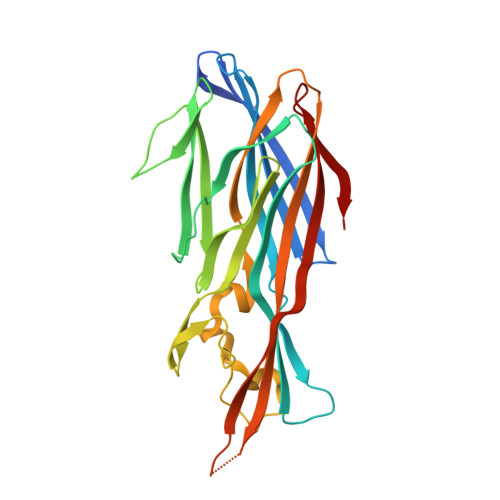
Panton-Valentine leukocidin (PVL), a bicomponent staphylococcal leukotoxin, is involved in the poor prognosis of necrotizing pneumonia. The present study aimed to elucidate the binding mechanism of PVL and in particular its cell-binding domain. The class S component of PVL, LukS-PV, is known to ensure cell targeting and exhibits the highest affinity for the neutrophil membrane (Kd∼10(-10) M) compared to the class F component of PVL, LukF-PV (Kd∼10(-9) M). Alanine scanning mutagenesis was used to identify the residues involved in LukS-PV binding to the neutrophil surface. Nineteen single alanine mutations were performed in the rim domain previously described as implicated in cell membrane interactions. Positions were chosen in order to replace polar or exposed charged residues and according to conservation between leukotoxin class S components. Characterization studies enabled to identify a cluster of residues essential for LukS-PV binding, localized on two loops of the rim domain. The mutations R73A, Y184A, T244A, H245A and Y250A led to dramatically reduced binding affinities for both human leukocytes and undifferentiated U937 cells expressing the C5a receptor. The three-dimensional structure of five of the mutants was determined using X-ray crystallography. Structure analysis identified residues Y184 and Y250 as crucial in providing structural flexibility in the receptor-binding domain of LukS-PV.
Organizational Affiliation:
Université de Strasbourg-CHRU, Fédération de Médecine Translationnelle de Strasbourg, EA 7290 Virulence bactérienne précoce, Institut de Bactériologie, Strasbourg, France.