Differential stabilities and sequence-dependent base pair opening dynamics of watson-crick base pairs with 5-hydroxymethylcytosine, 5-formylcytosine, or 5-carboxylcytosine.
Szulik, M.W., Pallan, P.S., Nocek, B., Voehler, M., Banerjee, S., Brooks, S., Joachimiak, A., Egli, M., Eichman, B.F., Stone, M.P.(2015) Biochemistry 54: 1294-1305
- PubMed: 25632825
- DOI: https://doi.org/10.1021/bi501534x
- Primary Citation of Related Structures:
4I9V, 4PWM, 4QC7 - PubMed Abstract:
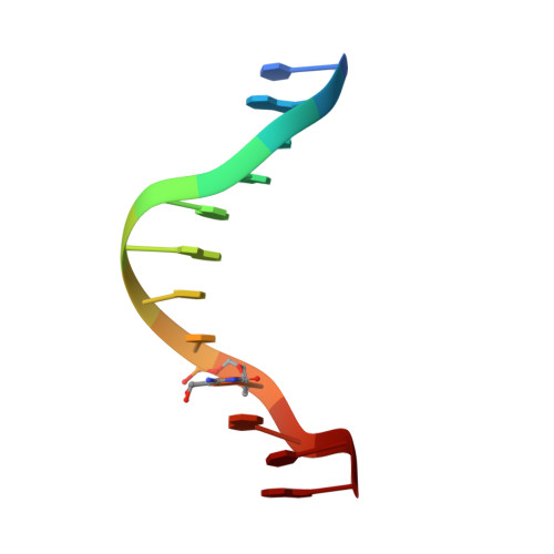
5-Hydroxymethylcytosine (5hmC), 5-formylcytosine (5fC), and 5-carboxylcytosine (5caC) form during active demethylation of 5-methylcytosine (5mC) and are implicated in epigenetic regulation of the genome. They are differentially processed by thymine DNA glycosylase (TDG), an enzyme involved in active demethylation of 5mC. Three modified Dickerson-Drew dodecamer (DDD) sequences, amenable to crystallographic and spectroscopic analyses and containing the 5'-CG-3' sequence associated with genomic cytosine methylation, containing 5hmC, 5fC, or 5caC placed site-specifically into the 5'-T(8)X(9)G(10)-3' sequence of the DDD, were compared. The presence of 5caC at the X(9) base increased the stability of the DDD, whereas 5hmC or 5fC did not. Both 5hmC and 5fC increased imino proton exchange rates and calculated rate constants for base pair opening at the neighboring base pair A(5):T(8), whereas 5caC did not. At the oxidized base pair G(4):X(9), 5fC exhibited an increase in the imino proton exchange rate and the calculated kop. In all cases, minimal effects to imino proton exchange rates occurred at the neighboring base pair C(3):G(10). No evidence was observed for imino tautomerization, accompanied by wobble base pairing, for 5hmC, 5fC, or 5caC when positioned at base pair G(4):X(9); each favored Watson-Crick base pairing. However, both 5fC and 5caC exhibited intranucleobase hydrogen bonding between their formyl or carboxyl oxygens, respectively, and the adjacent cytosine N(4) exocyclic amines. The lesion-specific differences observed in the DDD may be implicated in recognition of 5hmC, 5fC, or 5caC in DNA by TDG. However, they do not correlate with differential excision of 5hmC, 5fC, or 5caC by TDG, which may be mediated by differences in transition states of the enzyme-bound complexes.
Organizational Affiliation:
Department of Chemistry, Vanderbilt Institute of Chemical Biology, Vanderbilt Ingram Cancer Center, and Center for Structural Biology, Vanderbilt University , Nashville, Tennessee 37235, United States.