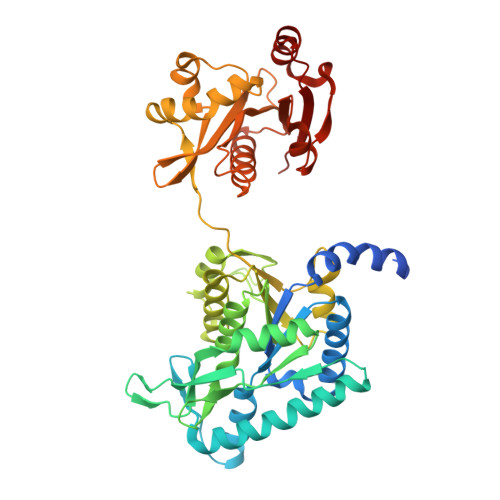
4I49

Structure of ngNAGS bound with bisubstrate analog CoA-NAG


Experimental Data Snapshot

  • Method: X-RAY DIFFRACTION
  • Resolution: 2.75 Å
  • R-Value Free: 0.271 
  • R-Value Work: 0.189 
  • R-Value Observed: 0.198 

wwPDB Validation   3D Report Full Report


Ligand Structure Quality Assessment 


This is version 1.3 of the entry. See complete history


Literature

Structure of the complex of Neisseria gonorrhoeae N-acetyl-l-glutamate synthase with a bound bisubstrate analog.

Zhao, G.Allewell, N.M.Tuchman, M.Shi, D.

(2013) Biochem Biophys Res Commun 430: 1253-1258

  • DOI: https://doi.org/10.1016/j.bbrc.2012.12.064

  • PubMed Abstract: 

    N-Acetyl-L-glutamate synthase catalyzes the conversion of AcCoA and glutamate to CoA and N-acetyl-L-glutamate (NAG), the first step of the arginine biosynthetic pathway in lower organisms. In mammals, NAG is an obligate cofactor of carbamoyl phosphate synthetase I in the urea cycle. We have previously reported the structures of NAGS from Neisseria gonorrhoeae (ngNAGS) with various substrates bound. Here we reported the preparation of the bisubstrate analog, CoA-S-acetyl-L-glutamate, the crystal structure of ngNAGS with CoA-NAG bound, and kinetic studies of several active site mutants. The results are consistent with a one-step nucleophilic addition-elimination mechanism with Glu353 as the catalytic base and Ser392 as the catalytic acid. The structure of the ngNAGS-bisubstrate complex together with the previous ngNAGS structures delineates the catalytic reaction path for ngNAGS.


  • Organizational Affiliation

    Center for Genetic Medicine Research and Department of Integrative Systems Biology, Children's National Medical Center, The George Washington University, Washington, DC 20010, USA.


Macromolecules
Find similar proteins by:  (by identity cutoff)  |  3D Structure
Entity ID: 1
MoleculeChains Sequence LengthOrganismDetailsImage
Amino-acid acetyltransferase456Neisseria gonorrhoeae SK-93-1035Mutation(s): 0 
Gene Names: argANGLG_00316
EC: 2.3.1.1
Entity Groups  
Sequence Clusters30% Identity50% Identity70% Identity90% Identity95% Identity100% Identity
Sequence Annotations
Expand
  • Reference Sequence
Small Molecules
Ligands 2 Unique
IDChains Name / Formula / InChI Key2D Diagram3D Interactions
1C4
Query on 1C4

Download Ideal Coordinates CCD File 
B [auth A](2S)-2-({(3S,5R,9R)-1-[(2R,3S,4R,5R)-5-(6-amino-9H-purin-9-yl)-4-hydroxy-3-(phosphonooxy)tetrahydrofuran-2-yl]-3,5,9-trihydroxy-8,8-dimethyl-3,5-dioxido-10,14,20-trioxo-2,4,6-trioxa-18-thia-11,15-diaza-3lambda~5~,5lambda~5~-diphosphaicosan-20-yl}amino)pentanedioic acid (non-preferred name)
C28 H45 N8 O21 P3 S
WCWCYRFADJEDNS-NOTSHUFBSA-N
SO4
Query on SO4

Download Ideal Coordinates CCD File 
C [auth A]SULFATE ION
O4 S
QAOWNCQODCNURD-UHFFFAOYSA-L
Experimental Data & Validation

Experimental Data

  • Method: X-RAY DIFFRACTION
  • Resolution: 2.75 Å
  • R-Value Free: 0.271 
  • R-Value Work: 0.189 
  • R-Value Observed: 0.198 
  • Space Group: P 3 1 2
Unit Cell:
Length ( Å )Angle ( ˚ )
a = 98.521α = 90
b = 98.521β = 90
c = 90.009γ = 120
Software Package:
Software NamePurpose
DENZOdata reduction
SCALEPACKdata scaling
PHENIXrefinement
PDB_EXTRACTdata extraction
StructureStudiodata collection

Structure Validation

View Full Validation Report


Ligand Structure Quality Assessment 


Entry History 

Deposition Data

Revision History  (Full details and data files)

  • Version 1.0: 2013-01-09
    Type: Initial release
  • Version 1.1: 2013-03-06
    Changes: Database references
  • Version 1.2: 2017-11-15
    Changes: Refinement description
  • Version 1.3: 2023-09-20
    Changes: Data collection, Database references, Derived calculations, Refinement description