The Arabidopsis B3 Domain Protein VERNALIZATION1 (VRN1) Is Involved in Processes Essential for Development, with Structural and Mutational Studies Revealing Its DNA-binding Surface.
King, G.J., Chanson, A.H., McCallum, E.J., Ohme-Takagi, M., Byriel, K., Hill, J.M., Martin, J.L., Mylne, J.S.(2013) J Biol Chem 288: 3198-3207
- PubMed: 23255593
- DOI: https://doi.org/10.1074/jbc.M112.438572
- Primary Citation of Related Structures:
4I1K - PubMed Abstract:
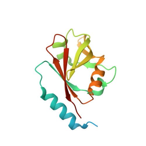
The B3 DNA-binding domain is a plant-specific domain found throughout the plant kingdom from the alga Chlamydomonas to grasses and flowering plants. Over 100 B3 domain-containing proteins are found in the model plant Arabidopsis thaliana, and one of these is critical for accelerating flowering in response to prolonged cold treatment, an epigenetic process called vernalization. Despite the specific phenotype of genetic vrn1 mutants, the VERNALIZATION1 (VRN1) protein localizes throughout the nucleus and shows sequence-nonspecific binding in vitro. In this work, we used a dominant repressor tag that overcomes genetic redundancy to show that VRN1 is involved in processes beyond vernalization that are essential for Arabidopsis development. To understand its sequence-nonspecific binding, we crystallized VRN1(208-341) and solved its crystal structure to 1.6 Å resolution using selenium/single-wavelength anomalous diffraction methods. The crystallized construct comprises the second VRN1 B3 domain and a preceding region conserved among VRN1 orthologs but absent in other B3 domains. We established the DNA-binding face using NMR and then mutated positively charged residues on this surface with a series of 16 Ala and Glu substitutions, ensuring that the protein fold was not disturbed using heteronuclear single quantum correlation NMR spectra. The triple mutant R249E/R289E/R296E was almost completely incapable of DNA binding in vitro. Thus, we have revealed that although VRN1 is sequence-nonspecific in DNA binding, it has a defined DNA-binding surface.
Organizational Affiliation:
Institute for Molecular Bioscience, University of Queensland, St Lucia, Brisbane, Queensland 4072, Australia.