4HDD

Domain swapping in the cytoplasmic domain of the Escherichia coli rhomboid protease


Experimental Data Snapshot

  • Method: X-RAY DIFFRACTION
  • Resolution: 1.35 Å
  • R-Value Free: 0.182 
  • R-Value Work: 0.162 
  • R-Value Observed: 0.163 

wwPDB Validation   3D Report Full Report


This is version 1.1 of the entry. See complete history


Literature

Domain swapping in the cytoplasmic domain of the Escherichia coli rhomboid protease.

Lazareno-Saez, C.Arutyunova, E.Coquelle, N.Lemieux, M.J.

(2013) J Mol Biol 425: 1127-1142

  • DOI: https://doi.org/10.1016/j.jmb.2013.01.019
  • Primary Citation of Related Structures:  
    4HDD

  • PubMed Abstract: 

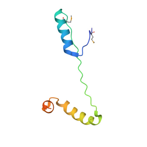
    Rhomboids are membrane-embedded serine proteases that cleave membrane protein substrates. Escherichia coli rhomboid GlpG (ecGlpG) consists of an N-terminal cytoplasmic domain and a membrane domain containing the active site. We determined the crystal structure of the soluble cytoplasmic domain of ecGlpG at 1.35Å resolution and examined whether this domain affected the catalytic activity of the enzyme. The structure revealed that the ecGlpG cytoplasmic domain exists as a dimer with extensive domain swapping between the two monomers. Domain-swapped dimers can be isolated from the full-length protein, suggesting that this is a physiologically relevant structure. An extensive steady-state kinetic analysis of the full-length ecGlpG and its membrane domain using soluble and transmembrane model protein substrates resulted in an unexpected conclusion: removal of the cytoplasmic domain does not alter the catalytic parameters for detergent-solubilized rhomboid for both substrates.


  • Organizational Affiliation

    Membrane Protein Disease Research Group, Department of Biochemistry, Faculty of Medicine and Dentistry, University of Alberta, Edmonton, Alberta, Canada T6G 2H7.


Macromolecules
Find similar proteins by:  (by identity cutoff)  |  3D Structure
Entity ID: 1
MoleculeChains Sequence LengthOrganismDetailsImage
Rhomboid protease GlpG79Escherichia coliMutation(s): 0 
Gene Names: b3424glpGJW5687
EC: 3.4.21.105
UniProt
Find proteins for P09391 (Escherichia coli (strain K12))
Explore P09391 
Go to UniProtKB:  P09391
Entity Groups  
Sequence Clusters30% Identity50% Identity70% Identity90% Identity95% Identity100% Identity
UniProt GroupP09391
Sequence Annotations
Expand
  • Reference Sequence
Small Molecules
Ligands 1 Unique
IDChains Name / Formula / InChI Key2D Diagram3D Interactions
ACT
Query on ACT

Download Ideal Coordinates CCD File 
B [auth A]ACETATE ION
C2 H3 O2
QTBSBXVTEAMEQO-UHFFFAOYSA-M
Modified Residues  1 Unique
IDChains TypeFormula2D DiagramParent
MSE
Query on MSE
A
L-PEPTIDE LINKINGC5 H11 N O2 SeMET
Experimental Data & Validation

Experimental Data

  • Method: X-RAY DIFFRACTION
  • Resolution: 1.35 Å
  • R-Value Free: 0.182 
  • R-Value Work: 0.162 
  • R-Value Observed: 0.163 
  • Space Group: I 4 2 2
Unit Cell:
Length ( Å )Angle ( ˚ )
a = 51.76α = 90
b = 51.76β = 90
c = 91.56γ = 90
Software Package:
Software NamePurpose
XSCALEdata scaling
PHENIXrefinement
PDB_EXTRACTdata extraction
MxDCdata collection
XDSdata reduction
PHENIXphasing

Structure Validation

View Full Validation Report


Entry History 

Deposition Data

Revision History  (Full details and data files)

  • Version 1.0: 2013-02-06
    Type: Initial release
  • Version 1.1: 2013-05-22
    Changes: Database references