4GX8

Crystal structure of a DNA polymerase III alpha-epsilon chimera


Experimental Data Snapshot

  • Method: X-RAY DIFFRACTION
  • Resolution: 1.70 Å
  • R-Value Free: 0.246 
  • R-Value Work: 0.213 
  • R-Value Observed: 0.214 

wwPDB Validation   3D Report Full Report


This is version 1.4 of the entry. See complete history


Literature

Proofreading exonuclease on a tether: the complex between the E. coli DNA polymerase III subunits alpha, {varepsilon}, theta and beta reveals a highly flexible arrangement of the proofreading domain

Ozawa, K.Horan, N.P.Robinson, A.Yagi, H.Hill, F.R.Jergic, S.Xu, Z.Q.Loscha, K.V.Li, N.Tehei, M.Oakley, A.J.Otting, G.Huber, T.Dixon, N.E.

(2013) Nucleic Acids Res 41: 5354-5367

  • DOI: https://doi.org/10.1093/nar/gkt162
  • Primary Citation of Related Structures:  
    4GX8, 4GX9

  • PubMed Abstract: 

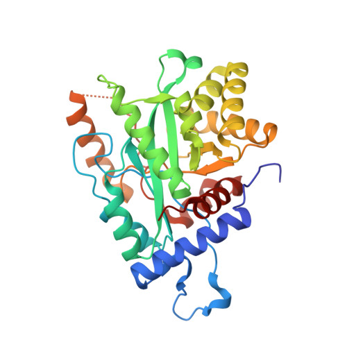
    A complex of the three (αεθ) core subunits and the β2 sliding clamp is responsible for DNA synthesis by Pol III, the Escherichia coli chromosomal DNA replicase. The 1.7 Å crystal structure of a complex between the PHP domain of α (polymerase) and the C-terminal segment of ε (proofreading exonuclease) subunits shows that ε is attached to α at a site far from the polymerase active site. Both α and ε contain clamp-binding motifs (CBMs) that interact simultaneously with β2 in the polymerization mode of DNA replication by Pol III. Strengthening of both CBMs enables isolation of stable αεθ:β2 complexes. Nuclear magnetic resonance experiments with reconstituted αεθ:β2 demonstrate retention of high mobility of a segment of 22 residues in the linker that connects the exonuclease domain of ε with its α-binding segment. In spite of this, small-angle X-ray scattering data show that the isolated complex with strengthened CBMs has a compact, but still flexible, structure. Photo-crosslinking with p-benzoyl-L-phenylalanine incorporated at different sites in the α-PHP domain confirm the conformational variability of the tether. Structural models of the αεθ:β2 replicase complex with primer-template DNA combine all available structural data.


  • Organizational Affiliation

    School of Chemistry, University of Wollongong, Northfields Avenue, Wollongong, NSW 2522, Australia.


Macromolecules
Find similar proteins by:  (by identity cutoff)  |  3D Structure
Entity ID: 1
MoleculeChains Sequence LengthOrganismDetailsImage
DNA polymerase III subunit epsilon,DNA polymerase III subunit alpha
A, B, C, D
315Escherichia coli K-12Mutation(s): 1 
Gene Names: dnaQmutDb0215JW0205dnaEpolCb0184JW0179
EC: 2.7.7.7
UniProt
Find proteins for P03007 (Escherichia coli (strain K12))
Explore P03007 
Go to UniProtKB:  P03007
Find proteins for P10443 (Escherichia coli (strain K12))
Explore P10443 
Go to UniProtKB:  P10443
Entity Groups  
Sequence Clusters30% Identity50% Identity70% Identity90% Identity95% Identity100% Identity
UniProt GroupsP03007P10443
Sequence Annotations
Expand
  • Reference Sequence
Experimental Data & Validation

Experimental Data

  • Method: X-RAY DIFFRACTION
  • Resolution: 1.70 Å
  • R-Value Free: 0.246 
  • R-Value Work: 0.213 
  • R-Value Observed: 0.214 
  • Space Group: P 1 21 1
Unit Cell:
Length ( Å )Angle ( ˚ )
a = 84.47α = 90
b = 56.63β = 93.52
c = 138.01γ = 90
Software Package:
Software NamePurpose
MAR345dtbdata collection
PHENIXmodel building
REFMACrefinement
MOSFLMdata reduction
SCALAdata scaling
PHENIXphasing

Structure Validation

View Full Validation Report


Entry History 

Deposition Data

Revision History  (Full details and data files)

  • Version 1.0: 2013-04-03
    Type: Initial release
  • Version 1.1: 2013-04-24
    Changes: Database references
  • Version 1.2: 2013-06-26
    Changes: Database references
  • Version 1.3: 2017-06-21
    Changes: Advisory
  • Version 1.4: 2023-11-08
    Changes: Data collection, Database references, Derived calculations, Refinement description