4GWN

Crystal structure of human mature meprin beta


Experimental Data Snapshot

  • Method: X-RAY DIFFRACTION
  • Resolution: 3.00 Å
  • R-Value Free: 0.238 
  • R-Value Work: 0.196 
  • R-Value Observed: 0.198 

wwPDB Validation   3D Report Full Report


Ligand Structure Quality Assessment 


This is version 2.1 of the entry. See complete history


Literature

Structural basis for the sheddase function of human meprin beta metalloproteinase at the plasma membrane

Arolas, J.L.Broder, C.Jefferson, T.Guevara, T.Sterchi, E.E.Bode, W.Stocker, W.Becker-Pauly, C.Gomis-Ruth, F.X.

(2012) Proc Natl Acad Sci U S A 109: 16131-16136

  • DOI: https://doi.org/10.1073/pnas.1211076109
  • Primary Citation of Related Structures:  
    4GWM, 4GWN

  • PubMed Abstract: 

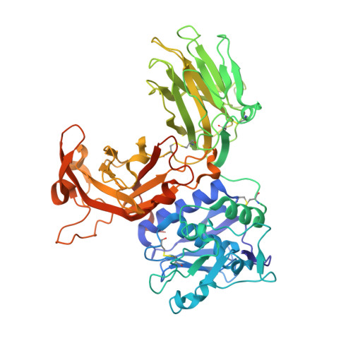
    Ectodomain shedding at the cell surface is a major mechanism to regulate the extracellular and circulatory concentration or the activities of signaling proteins at the plasma membrane. Human meprin β is a 145-kDa disulfide-linked homodimeric multidomain type-I membrane metallopeptidase that sheds membrane-bound cytokines and growth factors, thereby contributing to inflammatory diseases, angiogenesis, and tumor progression. In addition, it cleaves amyloid precursor protein (APP) at the β-secretase site, giving rise to amyloidogenic peptides. We have solved the X-ray crystal structure of a major fragment of the meprin β ectoprotein, the first of a multidomain oligomeric transmembrane sheddase, and of its zymogen. The meprin β dimer displays a compact shape, whose catalytic domain undergoes major rearrangement upon activation, and reveals an exosite and a sugar-rich channel, both of which possibly engage in substrate binding. A plausible structure-derived working mechanism suggests that substrates such as APP are shed close to the plasma membrane surface following an "N-like" chain trace.


  • Organizational Affiliation

    Proteolysis Laboratory, Department of Structural Biology, Molecular Biology Institute of Barcelona, Consejo Superior de Investigaciones Cientificas, Barcelona Science Park, E-08028 Barcelona, Spain.


Macromolecules
Find similar proteins by:  (by identity cutoff)  |  3D Structure
Entity ID: 1
MoleculeChains Sequence LengthOrganismDetailsImage
Meprin A subunit beta553Homo sapiensMutation(s): 0 
Gene Names: MEP1B
EC: 3.4.24.63
Membrane Entity: Yes 
UniProt & NIH Common Fund Data Resources
Find proteins for Q16820 (Homo sapiens)
Explore Q16820 
Go to UniProtKB:  Q16820
PHAROS:  Q16820
GTEx:  ENSG00000141434 
Entity Groups  
Sequence Clusters30% Identity50% Identity70% Identity90% Identity95% Identity100% Identity
UniProt GroupQ16820
Sequence Annotations
Expand
  • Reference Sequence
Oligosaccharides

Help

Entity ID: 2
MoleculeChains Length2D Diagram Glycosylation3D Interactions
2-acetamido-2-deoxy-beta-D-glucopyranose-(1-4)-2-acetamido-2-deoxy-beta-D-glucopyranose
B, E
2N-Glycosylation
Glycosylation Resources
GlyTouCan:  G42666HT
GlyCosmos:  G42666HT
GlyGen:  G42666HT
Entity ID: 3
MoleculeChains Length2D Diagram Glycosylation3D Interactions
alpha-D-mannopyranose-(1-3)-[alpha-D-mannopyranose-(1-6)]beta-D-mannopyranose-(1-4)-2-acetamido-2-deoxy-beta-D-glucopyranose-(1-4)-[alpha-L-fucopyranose-(1-3)][alpha-L-fucopyranose-(1-6)]2-acetamido-2-deoxy-beta-D-glucopyranose
C
7N-Glycosylation
Glycosylation Resources
GlyTouCan:  G11460AB
GlyCosmos:  G11460AB
GlyGen:  G11460AB
Entity ID: 4
MoleculeChains Length2D Diagram Glycosylation3D Interactions
alpha-D-mannopyranose-(1-2)-alpha-D-mannopyranose-(1-3)-[alpha-D-mannopyranose-(1-2)-alpha-D-mannopyranose-(1-6)]alpha-D-mannopyranose-(1-6)-[alpha-D-mannopyranose-(1-2)-alpha-D-mannopyranose-(1-3)]beta-D-mannopyranose-(1-4)-2-acetamido-2-deoxy-beta-D-glucopyranose-(1-4)-2-acetamido-2-deoxy-beta-D-glucopyranose
D
10N-Glycosylation
Glycosylation Resources
GlyTouCan:  G89864BN
GlyCosmos:  G89864BN
GlyGen:  G89864BN
Entity ID: 5
MoleculeChains Length2D Diagram Glycosylation3D Interactions
2-acetamido-2-deoxy-beta-D-glucopyranose-(1-4)-[alpha-L-fucopyranose-(1-6)]2-acetamido-2-deoxy-beta-D-glucopyranose
F
3N-Glycosylation
Glycosylation Resources
GlyTouCan:  G21290RB
GlyCosmos:  G21290RB
GlyGen:  G21290RB
Experimental Data & Validation

Experimental Data

  • Method: X-RAY DIFFRACTION
  • Resolution: 3.00 Å
  • R-Value Free: 0.238 
  • R-Value Work: 0.196 
  • R-Value Observed: 0.198 
  • Space Group: P 61 2 2
Unit Cell:
Length ( Å )Angle ( ˚ )
a = 74.96α = 90
b = 74.96β = 90
c = 502.65γ = 120
Software Package:
Software NamePurpose
ProDCdata collection
PHASERphasing
BUSTERrefinement
XDSdata reduction
SCALAdata scaling

Structure Validation

View Full Validation Report


Ligand Structure Quality Assessment 


Entry History 

Deposition Data

Revision History  (Full details and data files)

  • Version 1.0: 2012-09-19
    Type: Initial release
  • Version 1.1: 2012-10-31
    Changes: Database references
  • Version 1.2: 2017-11-15
    Changes: Refinement description
  • Version 2.0: 2020-07-29
    Type: Remediation
    Reason: Carbohydrate remediation
    Changes: Atomic model, Data collection, Derived calculations, Structure summary
  • Version 2.1: 2023-11-08
    Changes: Data collection, Database references, Refinement description, Structure summary