4GV4

Human ARTD3 (PARP3) - Catalytic domain in complex with inhibitor ME0328


Experimental Data Snapshot

  • Method: X-RAY DIFFRACTION
  • Resolution: 1.80 Å
  • R-Value Free: 0.203 
  • R-Value Work: 0.162 
  • R-Value Observed: 0.164 

wwPDB Validation   3D Report Full Report


Ligand Structure Quality Assessment 


This is version 1.2 of the entry. See complete history


Literature

PARP Inhibitor with Selectivity Toward ADP-Ribosyltransferase ARTD3/PARP3

Lindgren, A.E.G.Karlberg, T.Thorsell, A.G.Hesse, M.Spjut, S.Ekblad, T.Andersson, C.D.Pinto, A.F.Weigelt, J.Hottiger, M.O.Linusson, A.Elofsson, M.Schuler, H.

(2013) ACS Chem Biol 8: 1698-1703

  • DOI: https://doi.org/10.1021/cb4002014
  • Primary Citation of Related Structures:  
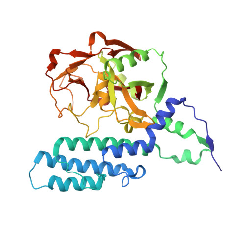
    4GV0, 4GV2, 4GV4, 4GV7

  • PubMed Abstract: 

    Inhibiting ADP-ribosyl transferases with PARP-inhibitors is considered a promising strategy for the treatment of many cancers and ischemia, but most of the cellular targets are poorly characterized. Here, we describe an inhibitor of ADP-ribosyltransferase-3/poly(ADP-ribose) polymerase-3 (ARTD3), a regulator of DNA repair and mitotic progression. In vitro profiling against 12 members of the enzyme family suggests selectivity for ARTD3, and crystal structures illustrate the molecular basis for inhibitor selectivity. The compound is active in cells, where it elicits ARTD3-specific effects at submicromolar concentration. Our results show that by targeting the nicotinamide binding site, selective inhibition can be achieved among the closest relatives of the validated clinical target, ADP-ribosyltransferase-1/poly(ADP-ribose) polymerase-1.


  • Organizational Affiliation

    Department of Chemistry, Umeå University, Umeå, Sweden.


Macromolecules
Find similar proteins by:  (by identity cutoff)  |  3D Structure
Entity ID: 1
MoleculeChains Sequence LengthOrganismDetailsImage
Poly [ADP-ribose] polymerase 3357Homo sapiensMutation(s): 0 
Gene Names: PARP3
EC: 2.4.2.30
UniProt & NIH Common Fund Data Resources
Find proteins for Q9Y6F1 (Homo sapiens)
Explore Q9Y6F1 
Go to UniProtKB:  Q9Y6F1
PHAROS:  Q9Y6F1
GTEx:  ENSG00000041880 
Entity Groups  
Sequence Clusters30% Identity50% Identity70% Identity90% Identity95% Identity100% Identity
UniProt GroupQ9Y6F1
Sequence Annotations
Expand
  • Reference Sequence
Small Molecules
Ligands 2 Unique
IDChains Name / Formula / InChI Key2D Diagram3D Interactions
MEJ
Query on MEJ

Download Ideal Coordinates CCD File 
B [auth A]3-(4-oxo-3,4-dihydroquinazolin-2-yl)-N-[(1S)-1-phenylethyl]propanamide
C19 H19 N3 O2
QIHBWVVVRYYYRO-ZDUSSCGKSA-N
DMS
Query on DMS

Download Ideal Coordinates CCD File 
C [auth A]DIMETHYL SULFOXIDE
C2 H6 O S
IAZDPXIOMUYVGZ-UHFFFAOYSA-N
Binding Affinity Annotations 
IDSourceBinding Affinity
MEJ Binding MOAD:  4GV4 IC50: 890 (nM) from 1 assay(s)
BindingDB:  4GV4 IC50: min: 890, max: 900 (nM) from 2 assay(s)
Experimental Data & Validation

Experimental Data

  • Method: X-RAY DIFFRACTION
  • Resolution: 1.80 Å
  • R-Value Free: 0.203 
  • R-Value Work: 0.162 
  • R-Value Observed: 0.164 
  • Space Group: P 1 21 1
Unit Cell:
Length ( Å )Angle ( ˚ )
a = 54.664α = 90
b = 56.742β = 112.74
c = 56.977γ = 90
Software Package:
Software NamePurpose
MxCuBEdata collection
MOLREPphasing
REFMACrefinement
XDSdata reduction
XSCALEdata scaling

Structure Validation

View Full Validation Report


Ligand Structure Quality Assessment 


Entry History 

Deposition Data

Revision History  (Full details and data files)

  • Version 1.0: 2013-06-19
    Type: Initial release
  • Version 1.1: 2013-10-16
    Changes: Database references
  • Version 1.2: 2023-11-08
    Changes: Data collection, Database references, Derived calculations, Refinement description