4GT1

Crystal structure of a MeaB- and MMAA-like GTPase from Mycobacterium tuberculosis bound to 2'-deoxyguanosine diphosphate


Experimental Data Snapshot

  • Method: X-RAY DIFFRACTION
  • Resolution: 2.00 Å
  • R-Value Free: 0.227 
  • R-Value Work: 0.189 
  • R-Value Observed: 0.191 

wwPDB Validation   3D Report Full Report


Ligand Structure Quality Assessment 


This is version 1.4 of the entry. See complete history


Literature

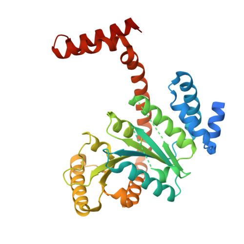
Crystal structures of Mycobacterial MeaB and MMAA-like GTPases.

Edwards, T.E.Baugh, L.Bullen, J.Baydo, R.O.Witte, P.Thompkins, K.Phan, I.Q.Abendroth, J.Clifton, M.C.Sankaran, B.Van Voorhis, W.C.Myler, P.J.Staker, B.L.Grundner, C.Lorimer, D.D.

(2015) J Struct Funct Genomics 16: 91-99

  • DOI: https://doi.org/10.1007/s10969-015-9197-2
  • Primary Citation of Related Structures:  
    3MD0, 3NXS, 3P32, 3TK1, 4GT1

  • PubMed Abstract: 

    The methylmalonyl Co-A mutase-associated GTPase MeaB from Methylobacterium extorquens is involved in glyoxylate regulation and required for growth. In humans, mutations in the homolog methylmalonic aciduria associated protein (MMAA) cause methylmalonic aciduria, which is often fatal. The central role of MeaB from bacteria to humans suggests that MeaB is also important in other, pathogenic bacteria such as Mycobacterium tuberculosis. However, the identity of the mycobacterial MeaB homolog is presently unclear. Here, we identify the M. tuberculosis protein Rv1496 and its homologs in M. smegmatis and M. thermoresistibile as MeaB. The crystal structures of all three homologs are highly similar to MeaB and MMAA structures and reveal a characteristic three-domain homodimer with GDP bound in the G domain active site. A structure of Rv1496 obtained from a crystal grown in the presence of GTP exhibited electron density for GDP, suggesting GTPase activity. These structures identify the mycobacterial MeaB and provide a structural framework for therapeutic targeting of M. tuberculosis MeaB.


  • Organizational Affiliation

    Beryllium, Seattle Structural Genomics Center for Infectious Disease (SSGCID), Bainbridge Island, WA, 98110, USA, tedwards@be4.com.


Macromolecules
Find similar proteins by:  (by identity cutoff)  |  3D Structure
Entity ID: 1
MoleculeChains Sequence LengthOrganismDetailsImage
Probable GTPase Rv1496/MT1543355Mycobacterium tuberculosisMutation(s): 0 
Gene Names: MT1543MTCY277.18Rv1496
EC: 3.6
UniProt
Find proteins for P9WPZ1 (Mycobacterium tuberculosis (strain ATCC 25618 / H37Rv))
Explore P9WPZ1 
Go to UniProtKB:  P9WPZ1
Entity Groups  
Sequence Clusters30% Identity50% Identity70% Identity90% Identity95% Identity100% Identity
UniProt GroupP9WPZ1
Sequence Annotations
Expand
  • Reference Sequence
Experimental Data & Validation

Experimental Data

Unit Cell:
Length ( Å )Angle ( ˚ )
a = 65.92α = 90
b = 187.82β = 90
c = 66.46γ = 90
Software Package:
Software NamePurpose
XSCALEdata scaling
PHASERphasing
REFMACrefinement
PDB_EXTRACTdata extraction
XDSdata reduction

Structure Validation

View Full Validation Report


Ligand Structure Quality Assessment 


Entry History 

Deposition Data

Revision History  (Full details and data files)

  • Version 1.0: 2012-09-05
    Type: Initial release
  • Version 1.1: 2015-03-11
    Changes: Database references
  • Version 1.2: 2015-04-15
    Changes: Database references
  • Version 1.3: 2015-06-03
    Changes: Database references
  • Version 1.4: 2023-09-13
    Changes: Data collection, Database references, Derived calculations, Refinement description