Change in single cystathionine beta-synthase domain-containing protein from a bent to flat conformation upon adenosine monophosphate binding
Jeong, B.C., Park, S.H., Yoo, K.S., Shin, J.S., Song, H.K.(2013) J Struct Biol 183: 40-46
- PubMed: 23664870
- DOI: https://doi.org/10.1016/j.jsb.2013.04.013
- Primary Citation of Related Structures:
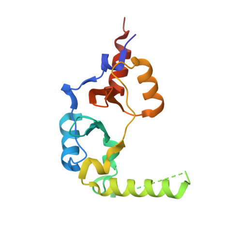
4GQY - PubMed Abstract:
Cystathionine β-synthase (CBS) domains are small intracellular modules that can act as binding domains for adenosine derivatives, and they may regulate the activity of associated enzymes or other functional domains. Among these, the single CBS domain-containing proteins, CBSXs, from Arabidopsis thaliana, have recently been identified as redox regulators of the thioredoxin system. Here, the crystal structure of CBSX2 in complex with adenosine monophosphate (AMP) is reported at 2.2Å resolution. The structure of dimeric CBSX2 with bound-AMP is shown to be approximately flat, which is in stark contrast to the bent form of apo-CBSXs. This conformational change in quaternary structure is triggered by a local structural change of the unique α5 helix, and by moving each loop P into an open conformation to accommodate incoming ligands. Furthermore, subtle rearrangement of the dimer interface triggers movement of all subunits, and consequently, the bent structure of the CBSX2 dimer becomes a flat structure. This reshaping of the structure upon complex formation with adenosine-containing ligand provides evidence that ligand-induced conformational reorganization of antiparallel CBS domains is an important regulatory mechanism.
Organizational Affiliation:
School of Life Sciences and Biotechnology, Korea University, Seoul 136-701, South Korea.