4GQB

Crystal Structure of the human PRMT5:MEP50 Complex


Experimental Data Snapshot

  • Method: X-RAY DIFFRACTION
  • Resolution: 2.06 Å
  • R-Value Free: 0.222 
  • R-Value Work: 0.188 
  • R-Value Observed: 0.190 

wwPDB Validation   3D Report Full Report


Ligand Structure Quality Assessment 


This is version 1.3 of the entry. See complete history


Literature

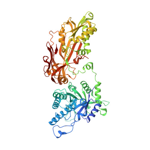
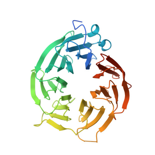
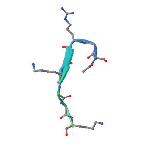
Crystal structure of the human PRMT5:MEP50 complex.

Antonysamy, S.Bonday, Z.Campbell, R.M.Doyle, B.Druzina, Z.Gheyi, T.Han, B.Jungheim, L.N.Qian, Y.Rauch, C.Russell, M.Sauder, J.M.Wasserman, S.R.Weichert, K.Willard, F.S.Zhang, A.Emtage, S.

(2012) Proc Natl Acad Sci U S A 109: 17960-17965

  • DOI: https://doi.org/10.1073/pnas.1209814109
  • Primary Citation of Related Structures:  
    4GQB

  • PubMed Abstract: 

    Protein arginine methyltransferases (PRMTs) play important roles in several cellular processes, including signaling, gene regulation, and transport of proteins and nucleic acids, to impact growth, differentiation, proliferation, and development. PRMT5 symmetrically di-methylates the two-terminal ω-guanidino nitrogens of arginine residues on substrate proteins. PRMT5 acts as part of a multimeric complex in concert with a variety of partner proteins that regulate its function and specificity. A core component of these complexes is the WD40 protein MEP50/WDR77/p44, which mediates interactions with binding partners and substrates. We have determined the crystal structure of human PRMT5 in complex with MEP50 (methylosome protein 50), bound to an S-adenosylmethionine analog and a peptide substrate derived from histone H4. The structure of the surprising hetero-octameric complex reveals the close interaction between the seven-bladed β-propeller MEP50 and the N-terminal domain of PRMT5, and delineates the structural elements of substrate recognition.


  • Organizational Affiliation

    Lilly Biotechnology Center, Eli Lilly and Company, San Diego, CA 92121, USA.


Macromolecules
Find similar proteins by:  (by identity cutoff)  |  3D Structure
Entity ID: 1
MoleculeChains Sequence LengthOrganismDetailsImage
Protein arginine N-methyltransferase 5637Homo sapiensMutation(s): 0 
Gene Names: PRMT5HRMT1L5IBP72JBP1SKB1
EC: 2.1.1 (PDB Primary Data), 2.1.1.125 (PDB Primary Data)
UniProt & NIH Common Fund Data Resources
Find proteins for O14744 (Homo sapiens)
Explore O14744 
Go to UniProtKB:  O14744
PHAROS:  O14744
GTEx:  ENSG00000100462 
Entity Groups  
Sequence Clusters30% Identity50% Identity70% Identity90% Identity95% Identity100% Identity
UniProt GroupO14744
Sequence Annotations
Expand
  • Reference Sequence
Find similar proteins by:  (by identity cutoff)  |  3D Structure
Entity ID: 2
MoleculeChains Sequence LengthOrganismDetailsImage
Methylosome protein 50344Homo sapiensMutation(s): 0 
Gene Names: WDR77MEP50HKMT1069Nbla10071
UniProt & NIH Common Fund Data Resources
Find proteins for Q9BQA1 (Homo sapiens)
Explore Q9BQA1 
Go to UniProtKB:  Q9BQA1
PHAROS:  Q9BQA1
GTEx:  ENSG00000116455 
Entity Groups  
Sequence Clusters30% Identity50% Identity70% Identity90% Identity95% Identity100% Identity
UniProt GroupQ9BQA1
Sequence Annotations
Expand
  • Reference Sequence

Find similar proteins by:  Sequence   |   3D Structure  

Entity ID: 3
MoleculeChains Sequence LengthOrganismDetailsImage
Histone H4 peptide22Homo sapiensMutation(s): 1 
UniProt & NIH Common Fund Data Resources
Find proteins for P62805 (Homo sapiens)
Explore P62805 
Go to UniProtKB:  P62805
PHAROS:  P62805
Entity Groups  
Sequence Clusters30% Identity50% Identity70% Identity90% Identity95% Identity100% Identity
UniProt GroupP62805
Sequence Annotations
Expand
  • Reference Sequence
Small Molecules
Ligands 1 Unique
IDChains Name / Formula / InChI Key2D Diagram3D Interactions
0XU
Query on 0XU

Download Ideal Coordinates CCD File 
D [auth A](2S,5S,6E)-2,5-diamino-6-[(3S,4R,5R)-5-(6-amino-9H-purin-9-yl)-3,4-dihydroxydihydrofuran-2(3H)-ylidene]hexanoic acid
C15 H21 N7 O5
YNKCTZQHSHSLNN-XVIGKWCNSA-N
Experimental Data & Validation

Experimental Data

  • Method: X-RAY DIFFRACTION
  • Resolution: 2.06 Å
  • R-Value Free: 0.222 
  • R-Value Work: 0.188 
  • R-Value Observed: 0.190 
  • Space Group: I 2 2 2
Unit Cell:
Length ( Å )Angle ( ˚ )
a = 101.981α = 90
b = 138.762β = 90
c = 178.472γ = 90
Software Package:
Software NamePurpose
BUSTERrefinement

Structure Validation

View Full Validation Report


Ligand Structure Quality Assessment 


Entry History 

Revision History  (Full details and data files)

  • Version 1.0: 2012-10-17
    Type: Initial release
  • Version 1.1: 2012-10-31
    Changes: Database references
  • Version 1.2: 2012-11-14
    Changes: Database references
  • Version 1.3: 2018-11-21
    Changes: Data collection, Structure summary