4GMW

Structure of rat cytosolic PEPCK Ld_1g in complex with PEP and GDP


Experimental Data Snapshot

  • Method: X-RAY DIFFRACTION
  • Resolution: 1.75 Å
  • R-Value Free: 0.259 
  • R-Value Work: 0.208 
  • R-Value Observed: 0.210 

wwPDB Validation   3D Report Full Report


Ligand Structure Quality Assessment 


This is version 1.3 of the entry. See complete history


Literature

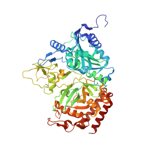
The {Omega}-loop lid domain of phosphoenolpyruvate carboxykinase is essential for catalytic function.

Johnson, T.A.Holyoak, T.

(2012) Biochemistry 51: 9547-9559

  • DOI: https://doi.org/10.1021/bi301278t
  • Primary Citation of Related Structures:  
    4GMM, 4GMU, 4GMW, 4GMZ, 4GNL, 4GNM, 4GNO, 4GNP, 4GNQ

  • PubMed Abstract: 

    Phosphoenolpyruvate carboxykinase (PEPCK) is an essential metabolic enzyme operating in the gluconeogenesis and glyceroneogenesis pathways. Recent studies have demonstrated that the enzyme contains a mobile active site lid domain that undergoes a transition between an open, disorded conformation and a closed, ordered conformation as the enzyme progresses through the catalytic cycle. The understanding of how this mobile domain functions in catalysis is incomplete. Previous studies showed that the closure of the lid domain stabilizes the reaction intermediate and protects the reactive intermediate from spurious protonation and thus contributes to the fidelity of the enzyme. To more fully investigate the roles of the lid domain in PEPCK function, we introduced three mutations that replaced the 11-residue lid domain with one, two, and three glycine residues. Kinetic analysis of the mutant enzymes demonstrates that none of the enzyme constructs exhibit any measurable kinetic activity, resulting in a decrease in the catalytic parameters of at least 10(6). Structural characterization of the mutants in complexes representing the catalytic cycle suggests that the inactivity is due to a role for the lid domain in the formation of the fully closed state of the enzyme that is required for catalytic function. In the absence of the lid domain, the enzyme is unable to achieve the fully closed state and is rendered inactive despite possessing all of the residues and substrates required for catalytic function. This work demonstrates how enzyme catalytic function can be abolished through the alteration of conformational equilibria despite all the elements required for chemical conversion of substrates to products remaining intact.


  • Organizational Affiliation

    Department of Biochemistry and Molecular Biology, The University of Kansas Medical Center, Kansas City, KS 66160, USA.


Macromolecules
Find similar proteins by:  (by identity cutoff)  |  3D Structure
Entity ID: 1
MoleculeChains Sequence LengthOrganismDetailsImage
Phosphoenolpyruvate carboxykinase, cytosolic [GTP]612Rattus norvegicusMutation(s): 0 
Gene Names: Pck1
EC: 4.1.1.32
UniProt
Find proteins for P07379 (Rattus norvegicus)
Explore P07379 
Go to UniProtKB:  P07379
Entity Groups  
Sequence Clusters30% Identity50% Identity70% Identity90% Identity95% Identity100% Identity
UniProt GroupP07379
Sequence Annotations
Expand
  • Reference Sequence
Small Molecules
Ligands 4 Unique
IDChains Name / Formula / InChI Key2D Diagram3D Interactions
GDP
Query on GDP

Download Ideal Coordinates CCD File 
B [auth A]GUANOSINE-5'-DIPHOSPHATE
C10 H15 N5 O11 P2
QGWNDRXFNXRZMB-UUOKFMHZSA-N
PEP
Query on PEP

Download Ideal Coordinates CCD File 
E [auth A]PHOSPHOENOLPYRUVATE
C3 H5 O6 P
DTBNBXWJWCWCIK-UHFFFAOYSA-N
MN
Query on MN

Download Ideal Coordinates CCD File 
C [auth A],
F [auth A],
G [auth A],
H [auth A]
MANGANESE (II) ION
Mn
WAEMQWOKJMHJLA-UHFFFAOYSA-N
NA
Query on NA

Download Ideal Coordinates CCD File 
D [auth A]SODIUM ION
Na
FKNQFGJONOIPTF-UHFFFAOYSA-N
Binding Affinity Annotations 
IDSourceBinding Affinity
PEP BindingDB:  4GMW Ki: 2.89e+5 (nM) from 1 assay(s)
Experimental Data & Validation

Experimental Data

  • Method: X-RAY DIFFRACTION
  • Resolution: 1.75 Å
  • R-Value Free: 0.259 
  • R-Value Work: 0.208 
  • R-Value Observed: 0.210 
  • Space Group: P 21 21 21
Unit Cell:
Length ( Å )Angle ( ˚ )
a = 61.091α = 90
b = 84.94β = 90
c = 119.322γ = 90
Software Package:
Software NamePurpose
SCALEPACKdata scaling
MOLREPphasing
REFMACrefinement
PDB_EXTRACTdata extraction
HKL-2000data collection
DENZOdata reduction

Structure Validation

View Full Validation Report


Ligand Structure Quality Assessment 


Entry History 

Deposition Data

Revision History  (Full details and data files)

  • Version 1.0: 2013-07-03
    Type: Initial release
  • Version 1.1: 2017-07-26
    Changes: Refinement description, Source and taxonomy
  • Version 1.2: 2017-11-15
    Changes: Refinement description
  • Version 1.3: 2023-09-13
    Changes: Data collection, Database references, Derived calculations, Refinement description