4GHF

Structure of Y257F variant of Homoprotocatechuate 2,3-Dioxygenase from B.fuscum in complex with 4-Nitrocatechol and dioxygen at 1.67 Ang resolution


Experimental Data Snapshot

  • Method: X-RAY DIFFRACTION
  • Resolution: 1.67 Å
  • R-Value Free: 0.181 
  • R-Value Work: 0.151 
  • R-Value Observed: 0.152 

wwPDB Validation   3D Report Full Report


Ligand Structure Quality Assessment 


This is version 1.2 of the entry. See complete history


Literature

Structural basis for the role of tyrosine 257 of homoprotocatechuate 2,3-dioxygenase in substrate and oxygen activation.

Kovaleva, E.G.Lipscomb, J.D.

(2012) Biochemistry 51: 8755-8763

  • DOI: https://doi.org/10.1021/bi301115c
  • Primary Citation of Related Structures:  
    4GHC, 4GHD, 4GHE, 4GHF, 4GHG, 4GHH

  • PubMed Abstract: 

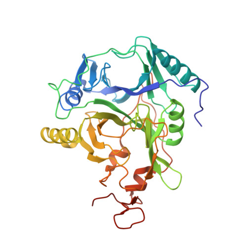
    Homoprotocatechuate 2,3-dioxygenase (FeHPCD) utilizes an active site Fe(II) to activate O(2) in a reaction cycle that ultimately results in aromatic ring cleavage. Here, the roles of the conserved active site residue Tyr257 are investigated by solving the X-ray crystal structures of the Tyr257-to-Phe variant (Y257F) in complex with the substrate homoprotocatechuate (HPCA) and the alternative substrate 4-nitrocatechol (4NC). These are compared with structures of the analogous wild type enzyme complexes. In addition, the oxy intermediate of the reaction cycle of Y257F-4NC + O(2) is formed in crystallo and structurally characterized. It is shown that both substrates adopt a previously unrecognized, slightly nonplanar, strained conformation affecting the geometries of all aromatic ring carbons when bound in the FeHPCD active site. This global deviation from planarity is not observed for the Y257F variant. In the Y257F-4NC-oxy complex, the O(2) is bound side-on to the Fe(II), while the 4NC is chelated in two adjacent sites. The ring of the 4NC in this complex is planar, in contrast to the equivalent FeHPCD intermediate, which exhibits substantial local distortion of the substrate hydroxyl moiety (C2-O(-)) that is hydrogen bonded to Tyr257. We propose that Tyr257 induces the global and local distortions of the substrate ring in two different ways. First, van der Waals conflict between the Tyr257-OH substituent and the substrate C2 carbon is relieved by adopting the globally strained structure. Second, Tyr257 stabilizes the localized out-of-plane position of the C2-O(-) by forming a stronger hydrogen bond as the distortion increases. Both types of distortions favor transfer of one electron out of the substrate to form a reactive semiquinone radical. Then, the localized distortion at substrate C2 promotes formation of the key alkylperoxo intermediate of the cycle resulting from oxygen attack on the activated substrate at C2, which becomes sp(3) hybridized. The inability of Y257F to promote the distorted substrate structure may explain the observed 100-fold decrease in the rates of the O(2) activation and insertion steps of the reaction.


  • Organizational Affiliation

    Institute of Molecular and Cellular Biology, University of Leeds, Leeds, LS2 9JT, UK. E.G.Kovaleva@leeds.ac.uk


Macromolecules
Find similar proteins by:  (by identity cutoff)  |  3D Structure
Entity ID: 1
MoleculeChains Sequence LengthOrganismDetailsImage
Homoprotocatechuate 2,3-dioxygenase
A, B, C, D
365Brevibacterium fuscumMutation(s): 1 
Gene Names: hpcd
EC: 1.13.11.15
UniProt
Find proteins for Q45135 (Brevibacterium fuscum)
Explore Q45135 
Go to UniProtKB:  Q45135
Entity Groups  
Sequence Clusters30% Identity50% Identity70% Identity90% Identity95% Identity100% Identity
UniProt GroupQ45135
Sequence Annotations
Expand
  • Reference Sequence
Small Molecules
Ligands 7 Unique
IDChains Name / Formula / InChI Key2D Diagram3D Interactions
P6G
Query on P6G

Download Ideal Coordinates CCD File 
G [auth A],
M [auth B],
N [auth B],
R [auth C],
W [auth D]
HEXAETHYLENE GLYCOL
C12 H26 O7
IIRDTKBZINWQAW-UHFFFAOYSA-N
PG4
Query on PG4

Download Ideal Coordinates CCD File 
L [auth B],
Q [auth C]
TETRAETHYLENE GLYCOL
C8 H18 O5
UWHCKJMYHZGTIT-UHFFFAOYSA-N
4NC
Query on 4NC

Download Ideal Coordinates CCD File 
F [auth A],
P [auth C],
V [auth D]
4-NITROCATECHOL
C6 H5 N O4
XJNPNXSISMKQEX-UHFFFAOYSA-N
FE2
Query on FE2

Download Ideal Coordinates CCD File 
E [auth A],
I [auth B],
O [auth C],
U [auth D]
FE (II) ION
Fe
CWYNVVGOOAEACU-UHFFFAOYSA-N
CA
Query on CA

Download Ideal Coordinates CCD File 
K [auth B]CALCIUM ION
Ca
BHPQYMZQTOCNFJ-UHFFFAOYSA-N
CL
Query on CL

Download Ideal Coordinates CCD File 
H [auth A],
J [auth B],
T [auth C],
Y [auth D]
CHLORIDE ION
Cl
VEXZGXHMUGYJMC-UHFFFAOYSA-M
OXY
Query on OXY

Download Ideal Coordinates CCD File 
S [auth C],
X [auth D]
OXYGEN MOLECULE
O2
MYMOFIZGZYHOMD-UHFFFAOYSA-N
Experimental Data & Validation

Experimental Data

  • Method: X-RAY DIFFRACTION
  • Resolution: 1.67 Å
  • R-Value Free: 0.181 
  • R-Value Work: 0.151 
  • R-Value Observed: 0.152 
  • Space Group: P 21 21 2
Unit Cell:
Length ( Å )Angle ( ˚ )
a = 110.519α = 90
b = 150.318β = 90
c = 96.105γ = 90
Software Package:
Software NamePurpose
REFMACrefinement
XDSdata reduction
SCALAdata scaling
REFMACphasing

Structure Validation

View Full Validation Report


Ligand Structure Quality Assessment 


Entry History 

Deposition Data

Revision History  (Full details and data files)

  • Version 1.0: 2012-10-31
    Type: Initial release
  • Version 1.1: 2012-12-12
    Changes: Database references
  • Version 1.2: 2023-09-13
    Changes: Data collection, Database references, Derived calculations, Refinement description