Structural basis for outer membrane sugar uptake in pseudomonads.
van den Berg, B.(2012) J Biol Chem 287: 41044-41052
- PubMed: 23066028
- DOI: https://doi.org/10.1074/jbc.M112.408518
- Primary Citation of Related Structures:
4GF4 - PubMed Abstract:
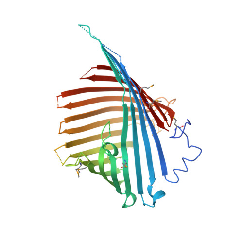
Substrate-specific outer membrane channels of gram-negative bacteria mediate uptake of many small molecules, including carbohydrates. The mechanism of sugar uptake by enterobacterial channels, such as Escherichia coli LamB (maltoporin), has been characterized in great detail. In pseudomonads and related organisms, sugar uptake is not mediated by LamB but by OprB channels. Beyond the notion that OprB channels seem to prefer monosaccharides as substrates, very little is known about OprB-mediated sugar uptake. Here I report the X-ray crystal structure of an OprB channel from Pseudomonas putida F1. The structure shows that OprB forms a monomeric, 16-stranded β-barrel with a constriction formed by extracellular loops L2 and L3. The side chains of two highly conserved arginine residues (Arg(83) and Arg(110)) and a conserved glutamate (Glu(106)) line the channel constriction and interact with a bound glucose molecule. Liposome swelling uptake assays show a strong preference for monosaccharide transport over disaccharides. Moreover, substrates with a net negative charge are disfavored by the channel, probably due to the negatively charged character of the constriction. The architecture of the eyelet and the absence of a greasy slide provide an explanation for the observed specificity of OprB for monosaccharides rather than the oligosaccharides preferred by LamB and related enterobacterial channels.
Organizational Affiliation:
Program in Molecular Medicine, University of Massachusetts Medical School, Worcester, Massachusetts 01605, USA. bert.vandenberg@umassmed.edu