4GDX

Crystal Structure of Human Gamma-Glutamyl Transpeptidase--Glutamate complex


Experimental Data Snapshot

  • Method: X-RAY DIFFRACTION
  • Resolution: 1.67 Å
  • R-Value Free: 0.174 
  • R-Value Work: 0.145 
  • R-Value Observed: 0.147 

wwPDB Validation   3D Report Full Report


Ligand Structure Quality Assessment 


This is version 1.4 of the entry. See complete history


Literature

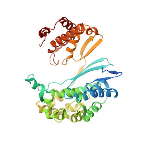
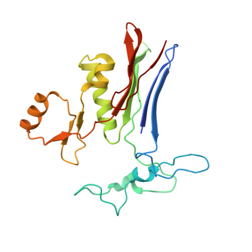
Novel Insights into Eukaryotic gamma-Glutamyltranspeptidase 1 from the Crystal Structure of the Glutamate-bound Human Enzyme.

West, M.B.Chen, Y.Wickham, S.Heroux, A.Cahill, K.Hanigan, M.H.Mooers, B.H.

(2013) J Biol Chem 288: 31902-31913

  • DOI: https://doi.org/10.1074/jbc.M113.498139
  • Primary Citation of Related Structures:  
    4GDX, 4GG2

  • PubMed Abstract: 

    The enzyme γ-glutamyltranspeptidase 1 (GGT1) is a conserved member of the N-terminal nucleophile hydrolase family that cleaves the γ-glutamyl bond of glutathione and other γ-glutamyl compounds. In animals, GGT1 is expressed on the surface of the cell and has critical roles in maintaining cysteine levels in the body and regulating intracellular redox status. Expression of GGT1 has been implicated as a potentiator of asthma, cardiovascular disease, and cancer. The rational design of effective inhibitors of human GGT1 (hGGT1) has been delayed by the lack of a reliable structural model. The available crystal structures of several bacterial GGTs have been of limited use due to differences in the catalytic behavior of bacterial and mammalian GGTs. We report the high resolution (1.67 Å) crystal structure of glutamate-bound hGGT1, the first of any eukaryotic GGT. Comparisons of the active site architecture of hGGT1 with those of its bacterial orthologs highlight key differences in the residues responsible for substrate binding, including a bimodal switch in the orientation of the catalytic nucleophile (Thr-381) that is unique to the human enzyme. Compared with several bacterial counterparts, the lid loop in the crystal structure of hGGT1 adopts an open conformation that allows greater access to the active site. The hGGT1 structure also revealed tightly bound chlorides near the catalytic residue that may contribute to catalytic activity. These are absent in the bacterial GGTs. These differences between bacterial and mammalian GGTs and the new structural data will accelerate the development of new therapies for GGT1-dependent diseases.


  • Organizational Affiliation

    From the Departments of Cell Biology and.


Macromolecules
Find similar proteins by:  (by identity cutoff)  |  3D Structure
Entity ID: 1
MoleculeChains Sequence LengthOrganismDetailsImage
Gamma-glutamyltranspeptidase 1 heavy chain374Homo sapiensMutation(s): 0 
Gene Names: GGTGGT1
EC: 2.3.2.2 (PDB Primary Data), 3.4.19.13 (PDB Primary Data), 3.4.19.14 (PDB Primary Data)
UniProt & NIH Common Fund Data Resources
Find proteins for P19440 (Homo sapiens)
Explore P19440 
Go to UniProtKB:  P19440
PHAROS:  P19440
GTEx:  ENSG00000100031 
Entity Groups  
Sequence Clusters30% Identity50% Identity70% Identity90% Identity95% Identity100% Identity
UniProt GroupP19440
Sequence Annotations
Expand
  • Reference Sequence
Find similar proteins by:  (by identity cutoff)  |  3D Structure
Entity ID: 2
MoleculeChains Sequence LengthOrganismDetailsImage
Gamma-glutamyltranspeptidase 1 light chain195Homo sapiensMutation(s): 0 
Gene Names: GGTGGT1
EC: 2.3.2.2 (PDB Primary Data), 3.4.19.13 (PDB Primary Data), 3.4.19.14 (PDB Primary Data)
UniProt & NIH Common Fund Data Resources
Find proteins for P19440 (Homo sapiens)
Explore P19440 
Go to UniProtKB:  P19440
PHAROS:  P19440
GTEx:  ENSG00000100031 
Entity Groups  
Sequence Clusters30% Identity50% Identity70% Identity90% Identity95% Identity100% Identity
UniProt GroupP19440
Sequence Annotations
Expand
  • Reference Sequence
Small Molecules
Ligands 4 Unique
IDChains Name / Formula / InChI Key2D Diagram3D Interactions
NAG
Query on NAG

Download Ideal Coordinates CCD File 
C [auth A]
D [auth A]
E [auth A]
F [auth A]
G [auth A]
C [auth A],
D [auth A],
E [auth A],
F [auth A],
G [auth A],
J [auth B]
2-acetamido-2-deoxy-beta-D-glucopyranose
C8 H15 N O6
OVRNDRQMDRJTHS-FMDGEEDCSA-N
GLU
Query on GLU

Download Ideal Coordinates CCD File 
K [auth B]GLUTAMIC ACID
C5 H9 N O4
WHUUTDBJXJRKMK-VKHMYHEASA-N
CL
Query on CL

Download Ideal Coordinates CCD File 
H [auth A],
L [auth B]
CHLORIDE ION
Cl
VEXZGXHMUGYJMC-UHFFFAOYSA-M
NA
Query on NA

Download Ideal Coordinates CCD File 
I [auth A]SODIUM ION
Na
FKNQFGJONOIPTF-UHFFFAOYSA-N
Experimental Data & Validation

Experimental Data

  • Method: X-RAY DIFFRACTION
  • Resolution: 1.67 Å
  • R-Value Free: 0.174 
  • R-Value Work: 0.145 
  • R-Value Observed: 0.147 
  • Space Group: C 2 2 21
Unit Cell:
Length ( Å )Angle ( ˚ )
a = 105.524α = 90
b = 125.247β = 90
c = 104.468γ = 90
Software Package:
Software NamePurpose
HKL-2000data collection
MOLREPphasing
PHENIXrefinement
HKL-2000data reduction
SCALAdata scaling

Structure Validation

View Full Validation Report


Ligand Structure Quality Assessment 


Entry History 

Deposition Data

Revision History  (Full details and data files)

  • Version 1.0: 2013-09-25
    Type: Initial release
  • Version 1.1: 2013-10-16
    Changes: Database references
  • Version 1.2: 2013-11-27
    Changes: Database references
  • Version 1.3: 2020-07-29
    Type: Remediation
    Reason: Carbohydrate remediation
    Changes: Data collection, Database references, Derived calculations, Structure summary
  • Version 1.4: 2023-09-13
    Changes: Data collection, Database references, Refinement description, Structure summary