


Crystal structure of Thioredoxin from Wuchereria bancrofti at 2.0 Angstrom
Nasser, Y., Prabhu, P., Ahmed, A., Iqbal, S., Betzel, C.To be published.
Experimental Data Snapshot
wwPDB Validation 3D Report Full Report

Entity ID: 1 | |||||
|---|---|---|---|---|---|
| Molecule | Chains | Sequence Length | Organism | Details | Image |
| Thioredoxin | 145 | Wuchereria bancrofti | Mutation(s): 0 Gene Names: TRX, trx-1 | 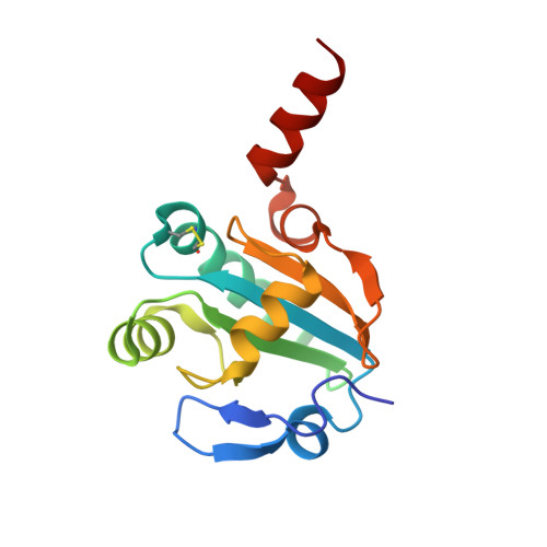 | |
UniProt | |||||
Find proteins for Q8ISA0 (Wuchereria bancrofti) Explore Q8ISA0 Go to UniProtKB: Q8ISA0 | |||||
Entity Groups | |||||
| Sequence Clusters | 30% Identity50% Identity70% Identity90% Identity95% Identity100% Identity | ||||
| UniProt Group | Q8ISA0 | ||||
Sequence AnnotationsExpand | |||||
| |||||
| Ligands 2 Unique | |||||
|---|---|---|---|---|---|
| ID | Chains | Name / Formula / InChI Key | 2D Diagram | 3D Interactions | |
| PEG Query on PEG | E [auth A], F [auth B], G [auth B] | DI(HYDROXYETHYL)ETHER C4 H10 O3 MTHSVFCYNBDYFN-UHFFFAOYSA-N |  | ||
| GOL Query on GOL | D [auth A] | GLYCEROL C3 H8 O3 PEDCQBHIVMGVHV-UHFFFAOYSA-N |  | ||
| Length ( Å ) | Angle ( ˚ ) |
|---|---|
| a = 112.61 | α = 90 |
| b = 112.61 | β = 90 |
| c = 162.05 | γ = 90 |
| Software Name | Purpose |
|---|---|
| DNA | data collection |
| REFMAC | refinement |
| MOSFLM | data reduction |
| SCALA | data scaling |
RCSB PDB (citation) is hosted by
RCSB PDB is a member of the

