


Crystal Structure of the Urokinase
Kang, Y.N., Stuckey, J.A., Nienaber, V., Giranda, V.To be published.
Experimental Data Snapshot
Entity ID: 1 | |||||
|---|---|---|---|---|---|
| Molecule | Chains | Sequence Length | Organism | Details | Image |
| Urokinase-type plasminogen activator | 246 | Homo sapiens | Mutation(s): 2 Gene Names: PLAU EC: 3.4.21.73 | 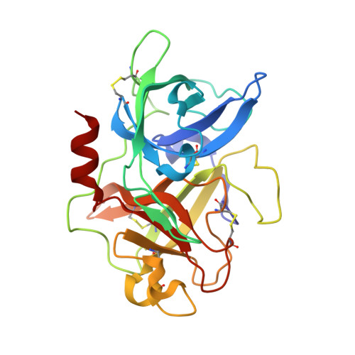 | |
UniProt & NIH Common Fund Data Resources | |||||
Find proteins for P00749 (Homo sapiens) Explore P00749 Go to UniProtKB: P00749 | |||||
PHAROS: P00749 GTEx: ENSG00000122861 | |||||
Entity Groups | |||||
| Sequence Clusters | 30% Identity50% Identity70% Identity90% Identity95% Identity100% Identity | ||||
| UniProt Group | P00749 | ||||
Sequence AnnotationsExpand | |||||
| |||||
| Ligands 3 Unique | |||||
|---|---|---|---|---|---|
| ID | Chains | Name / Formula / InChI Key | 2D Diagram | 3D Interactions | |
| 1UP Query on 1UP | F [auth A] | 2-[(7-carbamimidoyl-2-methoxynaphthalen-1-yl)oxy]acetamide C14 H15 N3 O3 ILLPJIZSGRJOPC-UHFFFAOYSA-N |  | ||
| SO4 Query on SO4 | B [auth A], C [auth A], D [auth A] | SULFATE ION O4 S QAOWNCQODCNURD-UHFFFAOYSA-L |  | ||
| ACT Query on ACT | E [auth A] | ACETATE ION C2 H3 O2 QTBSBXVTEAMEQO-UHFFFAOYSA-M |  | ||
| Length ( Å ) | Angle ( ˚ ) |
|---|---|
| a = 55.1 | α = 90 |
| b = 52.4 | β = 90 |
| c = 80.25 | γ = 90 |
| Software Name | Purpose |
|---|---|
| DENZO | data reduction |
| SCALEPACK | data scaling |
| BUSTER-TNT | refinement |
| PDB_EXTRACT | data extraction |
| BUSTER | refinement |
RCSB PDB (citation) is hosted by
RCSB PDB is a member of the

