Structural understanding of the glutathione-dependent reduction mechanism of glutathionyl-hydroquinone reductases.
Green, A.R., Hayes, R.P., Xun, L., Kang, C.(2012) J Biol Chem 287: 35838-35848
- PubMed: 22955277
- DOI: https://doi.org/10.1074/jbc.M112.395541
- Primary Citation of Related Structures:
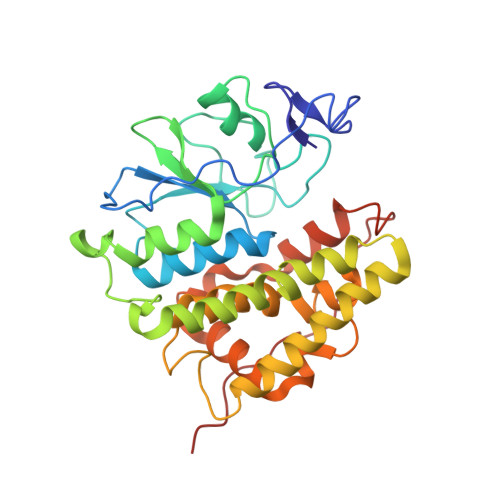
4FQU, 4G0I, 4G0K, 4G0L - PubMed Abstract:
Glutathionyl-hydroquinone reductases (GS- HQRs) are a newly identified group of glutathione transferases, and they are widely distributed in bacteria, halobacteria, fungi, and plants. GS-HQRs catalyze glutathione (GSH)-dependent reduction of glutathionyl-hydroquinones (GS-hydroquinones) to hydroquinones. GS-hydroquinones can be spontaneously formed from benzoquinones reacting with reduced GSH via Michael addition, and GS-HQRs convert the conjugates to hydroquinones. In this report we have determined the structures of two bacterial GS-HQRs, PcpF of Sphingobium chlorophenolicum and YqjG of Escherichia coli. The two structures and the previously reported structure of a fungal GS-HQR shared many features and displayed complete conservation for all the critical residues. Furthermore, we obtained the binary complex structures with GS-menadione, which in its reduced form, GS-menadiol, is a substrate. The structure revealed a large H-site that could accommodate various substituted hydroquinones and a hydrogen network of three Tyr residues that could provide the proton for reductive deglutathionylation. Mutation of the Tyr residues and the position of two GSH molecules confirmed the proposed mechanism of GS-HQRs. The conservation of GS-HQRs across bacteria, halobacteria, fungi, and plants potentiates the physiological role of these enzymes in quinone metabolism.
Organizational Affiliation:
School of Molecular Biosciences, Washington State University, Pullman, Washington 99164, USA.