4FH5

Crystal structures of the Cid1 poly (U) polymerase reveal the mechanism for UTP selectivity - MgUTP bound


Experimental Data Snapshot

  • Method: X-RAY DIFFRACTION
  • Resolution: 2.30 Å
  • R-Value Free: 0.244 
  • R-Value Work: 0.202 
  • R-Value Observed: 0.204 

wwPDB Validation   3D Report Full Report


Ligand Structure Quality Assessment 


This is version 1.2 of the entry. See complete history


Literature

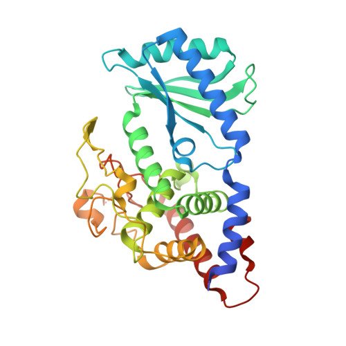
Crystal structures of the Cid1 poly (U) polymerase reveal the mechanism for UTP selectivity.

Lunde, B.M.Magler, I.Meinhart, A.

(2012) Nucleic Acids Res 40: 9815-9824

  • DOI: https://doi.org/10.1093/nar/gks740
  • Primary Citation of Related Structures:  
    4FH3, 4FH5, 4FHP, 4FHV, 4FHW, 4FHX, 4FHY

  • PubMed Abstract: 

    Polyuridylation is emerging as a ubiquitous post-translational modification with important roles in multiple aspects of RNA metabolism. These poly (U) tails are added by poly (U) polymerases with homology to poly (A) polymerases; nevertheless, the selection for UTP over ATP remains enigmatic. We report the structures of poly (U) polymerase Cid1 from Schizoscaccharomyces pombe alone and in complex with UTP, CTP, GTP and 3'-dATP. These structures reveal that each of the 4 nt can be accommodated at the active site; however, differences exist that suggest how the polymerase selects UTP over the other nucleotides. Furthermore, we find that Cid1 shares a number of common UTP recognition features with the kinetoplastid terminal uridyltransferases. Kinetic analysis of Cid1's activity for its preferred substrates, UTP and ATP, reveal a clear preference for UTP over ATP. Ultimately, we show that a single histidine in the active site plays a pivotal role for poly (U) activity. Notably, this residue is typically replaced by an asparagine residue in Cid1-family poly (A) polymerases. By mutating this histidine to an asparagine residue in Cid1, we diminished Cid1's activity for UTP addition and improved ATP incorporation, supporting that this residue is important for UTP selectivity.


  • Organizational Affiliation

    Department of Biomolecular Mechanisms, Max-Planck-Institute for Medical Research, Jahnstrasse 29, 69120 Heidelberg, Germany.


Macromolecules
Find similar proteins by:  (by identity cutoff)  |  3D Structure
Entity ID: 1
MoleculeChains Sequence LengthOrganismDetailsImage
Poly(A) RNA polymerase protein cid1349Schizosaccharomyces pombe 972h-Mutation(s): 0 
Gene Names: cid1SPAC19D5.03
EC: 2.7.7
UniProt
Find proteins for O13833 (Schizosaccharomyces pombe (strain 972 / ATCC 24843))
Explore O13833 
Go to UniProtKB:  O13833
Entity Groups  
Sequence Clusters30% Identity50% Identity70% Identity90% Identity95% Identity100% Identity
UniProt GroupO13833
Sequence Annotations
Expand
  • Reference Sequence
Small Molecules
Ligands 2 Unique
IDChains Name / Formula / InChI Key2D Diagram3D Interactions
UTP
Query on UTP

Download Ideal Coordinates CCD File 
B [auth A]URIDINE 5'-TRIPHOSPHATE
C9 H15 N2 O15 P3
PGAVKCOVUIYSFO-XVFCMESISA-N
MG
Query on MG

Download Ideal Coordinates CCD File 
C [auth A]MAGNESIUM ION
Mg
JLVVSXFLKOJNIY-UHFFFAOYSA-N
Experimental Data & Validation

Experimental Data

  • Method: X-RAY DIFFRACTION
  • Resolution: 2.30 Å
  • R-Value Free: 0.244 
  • R-Value Work: 0.202 
  • R-Value Observed: 0.204 
  • Space Group: P 21 21 21
Unit Cell:
Length ( Å )Angle ( ˚ )
a = 53.58α = 90
b = 62.86β = 90
c = 111.5γ = 90
Software Package:
Software NamePurpose
XDSdata scaling
REFMACrefinement
XDSdata reduction
REFMACphasing

Structure Validation

View Full Validation Report


Ligand Structure Quality Assessment 


Entry History 

Deposition Data

Revision History  (Full details and data files)

  • Version 1.0: 2012-08-29
    Type: Initial release
  • Version 1.1: 2012-11-07
    Changes: Database references
  • Version 1.2: 2023-09-13
    Changes: Data collection, Database references, Derived calculations, Refinement description