4FBZ

Crystal structure of deltarhodopsin from Haloterrigena thermotolerans


Experimental Data Snapshot

  • Method: X-RAY DIFFRACTION
  • Resolution: 2.70 Å
  • R-Value Free: 0.261 
  • R-Value Work: 0.235 
  • R-Value Observed: 0.235 

wwPDB Validation   3D Report Full Report


Ligand Structure Quality Assessment 


This is version 1.3 of the entry. See complete history


Literature

Crystal structure of deltarhodopsin-3 from Haloterrigena thermotolerans

Zhang, J.Mizuno, K.Murata, Y.Koide, H.Murakami, M.Ihara, K.Kouyama, T.

(2013) Proteins 81: 1585-1592

  • DOI: https://doi.org/10.1002/prot.24316
  • Primary Citation of Related Structures:  
    4FBZ

  • PubMed Abstract: 

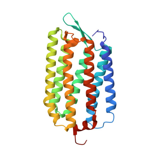
    Deltarhodopsin, a new member of the microbial rhodopsin family, functions as a light-driven proton pump. Here, we report the three-dimensional structure of deltarhodopsin (dR3) from Haloterrigena thermotolerans at 2.7 Å resolution. A crystal belonging to space group R32 (a, b = 111.71 Å, c = 198.25 Å) was obtained by the membrane fusion method. In this crystal, dR3 forms a trimeric structure as observed for bacteriorhodopsin (bR). Structural comparison of dR with bR showed that the inner part (the proton release and uptake pathways) is highly conserved. Meanwhile, residues in the protein-protein contact region are largely altered so that the diameter of the trimeric structure at the cytoplasmic side is noticeably larger in dR3. Unlike bR, dR3 possesses a helical segment at the C-terminal region that fills the space between the AB and EF loops. A significant difference is also seen in the FG loop, which is one residue longer in dR3. Another peculiar property of dR3 is a highly crowded distribution of positively charged residues on the cytoplasmic surface, which may be relevant to a specific interaction with some cytoplasmic component.


  • Organizational Affiliation

    Department of Physics, Graduate School of Science, Nagoya University, Nagoya, Japan.


Macromolecules
Find similar proteins by:  (by identity cutoff)  |  3D Structure
Entity ID: 1
MoleculeChains Sequence LengthOrganismDetailsImage
deltarhodopsin241Natrinema thermotoleransMutation(s): 0 
Membrane Entity: Yes 
UniProt
Find proteins for I4DST7 (Natrinema thermotolerans)
Explore I4DST7 
Go to UniProtKB:  I4DST7
Entity Groups  
Sequence Clusters30% Identity50% Identity70% Identity90% Identity95% Identity100% Identity
UniProt GroupI4DST7
Sequence Annotations
Expand
  • Reference Sequence
Small Molecules
Ligands 6 Unique
IDChains Name / Formula / InChI Key2D Diagram3D Interactions
22B
Query on 22B

Download Ideal Coordinates CCD File 
C [auth A]BACTERIORUBERIN
C50 H76 O4
UVCQMCCIAHQDAF-CUMPQFAQSA-N
L2P
Query on L2P

Download Ideal Coordinates CCD File 
E [auth A]2,3-DI-PHYTANYL-GLYCEROL
C43 H88 O3
ISDBCJSGCHUHFI-UMZPFTBHSA-N
SQL
Query on SQL

Download Ideal Coordinates CCD File 
D [auth A](6E,10E,14E,18E)-2,6,10,15,19,23-hexamethyltetracosa-2,6,10,14,18,22-hexaene
C30 H50
YYGNTYWPHWGJRM-AAJYLUCBSA-N
BNG
Query on BNG

Download Ideal Coordinates CCD File 
F [auth A],
G [auth A]
nonyl beta-D-glucopyranoside
C15 H30 O6
QFAPUKLCALRPLH-UXXRCYHCSA-N
RET
Query on RET

Download Ideal Coordinates CCD File 
B [auth A]RETINAL
C20 H28 O
NCYCYZXNIZJOKI-OVSJKPMPSA-N
SO4
Query on SO4

Download Ideal Coordinates CCD File 
H [auth A],
I [auth A],
J [auth A]
SULFATE ION
O4 S
QAOWNCQODCNURD-UHFFFAOYSA-L
Experimental Data & Validation

Experimental Data

  • Method: X-RAY DIFFRACTION
  • Resolution: 2.70 Å
  • R-Value Free: 0.261 
  • R-Value Work: 0.235 
  • R-Value Observed: 0.235 
  • Space Group: H 3 2
Unit Cell:
Length ( Å )Angle ( ˚ )
a = 111.71α = 90
b = 111.71β = 90
c = 198.25γ = 120
Software Package:
Software NamePurpose
ADSCdata collection
CNSrefinement
MOSFLMdata reduction
SCALAdata scaling
CNSphasing

Structure Validation

View Full Validation Report


Ligand Structure Quality Assessment 


Entry History 

Deposition Data

  • Released Date: 2013-05-15 
  • Deposition Author(s): Kouyama, T.

Revision History  (Full details and data files)

  • Version 1.0: 2013-05-15
    Type: Initial release
  • Version 1.1: 2013-10-09
    Changes: Database references
  • Version 1.2: 2020-07-29
    Type: Remediation
    Reason: Carbohydrate remediation
    Changes: Data collection, Derived calculations, Structure summary
  • Version 1.3: 2023-11-08
    Changes: Data collection, Database references, Refinement description, Structure summary