4F12

Crystal structure of the extracellular domain of human GABA(B) receptor GBR2


Experimental Data Snapshot

  • Method: X-RAY DIFFRACTION
  • Resolution: 3.02 Å
  • R-Value Free: 0.254 
  • R-Value Work: 0.207 
  • R-Value Observed: 0.209 

wwPDB Validation   3D Report Full Report


Ligand Structure Quality Assessment 


This is version 2.0 of the entry. See complete history


Literature

Structure and functional interaction of the extracellular domain of human GABA(B) receptor GBR2.

Geng, Y.Xiong, D.Mosyak, L.Malito, D.L.Kniazeff, J.Chen, Y.Burmakina, S.Quick, M.Bush, M.Javitch, J.A.Pin, J.P.Fan, Q.R.

(2012) Nat Neurosci 15: 970-978

  • DOI: https://doi.org/10.1038/nn.3133
  • Primary Citation of Related Structures:  
    4F11, 4F12

  • PubMed Abstract: 

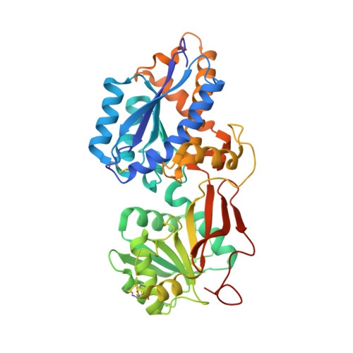
    Inhibitory neurotransmission is mediated primarily by GABA. The metabotropic GABA(B) receptor is a G protein-coupled receptor central to mammalian brain function. Malfunction of GABA(B) receptor has been implicated in several neurological disorders. GABA(B) receptor functions as a heterodimeric assembly of GBR1 and GBR2 subunits, where GBR1 is responsible for ligand-binding and GBR2 is responsible for G protein coupling. Here we demonstrate that the GBR2 ectodomain directly interacts with the GBR1 ectodomain to increase agonist affinity by selectively stabilizing the agonist-bound conformation of GBR1. We present the crystal structure of the GBR2 ectodomain, which reveals a polar heterodimeric interface. We also identify specific heterodimer contacts from both subunits, and GBR1 residues involved in ligand recognition. Lastly, our structural and functional data indicate that the GBR2 ectodomain adopts a constitutively open conformation, suggesting a structural asymmetry in the active state of GABA(B) receptor that is unique to the GABAergic system.


  • Organizational Affiliation

    Department of Pharmacology, Columbia University, New York, New York, USA.


Macromolecules
Find similar proteins by:  (by identity cutoff)  |  3D Structure
Entity ID: 1
MoleculeChains Sequence LengthOrganismDetailsImage
Gamma-aminobutyric acid type B receptor subunit 2433Homo sapiensMutation(s): 0 
Gene Names: GABBR2GPR51GPRC3B
UniProt & NIH Common Fund Data Resources
Find proteins for O75899 (Homo sapiens)
Explore O75899 
Go to UniProtKB:  O75899
PHAROS:  O75899
GTEx:  ENSG00000136928 
Entity Groups  
Sequence Clusters30% Identity50% Identity70% Identity90% Identity95% Identity100% Identity
UniProt GroupO75899
Sequence Annotations
Expand
  • Reference Sequence
Oligosaccharides

Help

Entity ID: 2
MoleculeChains Length2D Diagram Glycosylation3D Interactions
2-acetamido-2-deoxy-beta-D-glucopyranose-(1-4)-[alpha-L-fucopyranose-(1-6)]2-acetamido-2-deoxy-beta-D-glucopyranose
B
3N-Glycosylation
Glycosylation Resources
GlyTouCan:  G21290RB
GlyCosmos:  G21290RB
GlyGen:  G21290RB
Experimental Data & Validation

Experimental Data

  • Method: X-RAY DIFFRACTION
  • Resolution: 3.02 Å
  • R-Value Free: 0.254 
  • R-Value Work: 0.207 
  • R-Value Observed: 0.209 
  • Space Group: C 2 2 21
Unit Cell:
Length ( Å )Angle ( ˚ )
a = 82.684α = 90
b = 142.515β = 90
c = 79.264γ = 90
Software Package:
Software NamePurpose
DENZOdata reduction
SCALEPACKdata scaling
REFMACrefinement
PDB_EXTRACTdata extraction
HKL-2000data collection
HKL-2000data reduction
HKL-2000data scaling
PHASERphasing

Structure Validation

View Full Validation Report


Ligand Structure Quality Assessment 


Entry History 

Deposition Data

Revision History  (Full details and data files)

  • Version 1.0: 2012-06-06
    Type: Initial release
  • Version 1.1: 2012-08-15
    Changes: Database references
  • Version 1.2: 2017-11-15
    Changes: Refinement description
  • Version 2.0: 2020-07-29
    Type: Remediation
    Reason: Carbohydrate remediation
    Changes: Atomic model, Data collection, Database references, Derived calculations, Structure summary