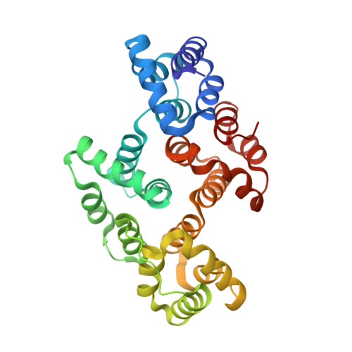
Alpha-1 giardin is an annexin with highly unusual calcium-regulated mechanisms
Weeratunga, S.K., Osman, A., Hu, N.-J., Wang, C.K., Mason, L., Svard, S., Hope, G., Jones, M.K., Hofmann, A.(2012) J Mol Biol 423: 169-181
- PubMed: 22796298
- DOI: https://doi.org/10.1016/j.jmb.2012.06.041
- Primary Citation of Related Structures:
4EVF, 4EVH - PubMed Abstract:
Alpha-giardins constitute the annexin proteome (group E annexins) in the intestinal protozoan parasite Giardia and, as such, represent the evolutionary oldest eukaryotic annexins. The dominance of alpha-giardins in the cytoskeleton of Giardia with its greatly reduced actin content emphasises the importance of the alpha-giardins for the structural integrity of the parasite, which is particularly critical in the transformation stage between cyst and trophozoite. In this study, we report the crystal structures of the apo- and calcium-bound forms of α1-giardin, a protein localised to the plasma membrane of Giardia trophozoites that has recently been identified as a vaccine target. The calcium-bound crystal structure of α1-giardin revealed the presence of a type III site in the first repeat as known from other annexin structures, as well as a novel calcium binding site situated between repeats I and IV. By means of comparison, the crystal structures of three different alpha-giardins known to date indicate that these proteins engage different calcium coordination schemes, among each other, as well as compared to annexins of groups A-D. Evaluation of the calcium-dependent binding to acidic phosphoplipid membranes revealed that this process is not only mediated but also regulated by the environmental calcium concentration. Uniquely within the large family of annexins, α1-giardin disengages from the phospholipid membrane at high calcium concentrations possibly due to formation of a dimeric species. The observed behaviour is in line with changing calcium levels experienced by the parasite during excystation and may thus provide first insights into the molecular mechanisms underpinning the transformation and survival of the parasite in the host.
Organizational Affiliation:
Structural Chemistry Program, Eskitis Institute for Cell and Molecular Therapies, Griffith University, Brisbane, Qld 4111, Australia.