4EUZ

Crystal structure of serratia fonticola carbapenemase SFC-1 S70A-Meropenem complex


Experimental Data Snapshot

  • Method: X-RAY DIFFRACTION
  • Resolution: 1.08 Å
  • R-Value Free: 0.138 
  • R-Value Work: 0.108 
  • R-Value Observed: 0.115 

wwPDB Validation   3D Report Full Report


Ligand Structure Quality Assessment 


This is version 1.2 of the entry. See complete history


Literature

The basis for carbapenem hydrolysis by class A beta-lactamases: a combined investigation using crystallography and simulations.

Fonseca, F.Chudyk, E.I.van der Kamp, M.W.Correia, A.Mulholland, A.J.Spencer, J.

(2012) J Am Chem Soc 134: 18275-18285

  • DOI: https://doi.org/10.1021/ja304460j
  • Primary Citation of Related Structures:  
    4EQI, 4EUZ, 4EV4

  • PubMed Abstract: 

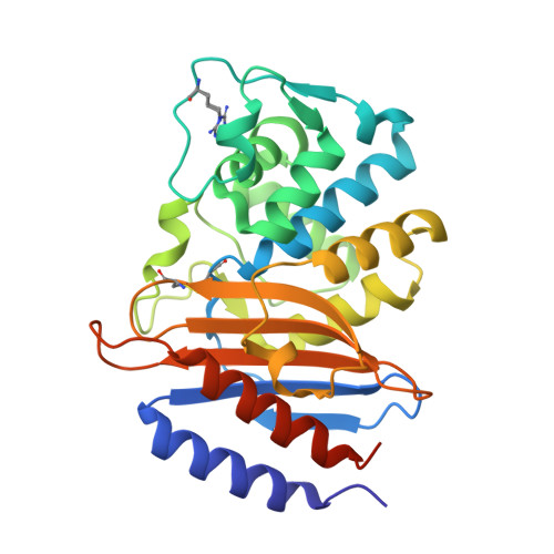
    Carbapenems are the most potent β-lactam antibiotics and key drugs for treating infections by Gram-negative bacteria. In such organisms, β-lactam resistance arises principally from β-lactamase production. Although carbapenems escape the activity of most β-lactamases, due in the class A enzymes to slow deacylation of the covalent acylenzyme intermediate, carbapenem-hydrolyzing class A β-lactamases are now disseminating in clinically relevant bacteria. The reasons why carbapenems are substrates for these enzymes, but inhibit other class A β-lactamases, remain to be fully established. Here, we present crystal structures of the class A carbapenemase SFC-1 from Serratia fonticola and of complexes of its Ser70 Ala (Michaelis) and Glu166 Ala (acylenzyme) mutants with the carbapenem meropenem. These are the first crystal structures of carbapenem complexes of a class A carbapenemase. Our data reveal that, in the SFC-1 acylenzyme complex, the meropenem 6α-1R-hydroxyethyl group interacts with Asn132, but not with the deacylating water molecule. Molecular dynamics simulations indicate that this mode of binding occurs in both the Michaelis and acylenzyme complexes of wild-type SFC-1. In carbapenem-inhibited class A β-lactamases, it is proposed that the deacylating water molecule is deactivated by interaction with the carbapenem 6α-1R-hydroxyethyl substituent. Structural comparisons with such enzymes suggest that in SFC-1 subtle repositioning of key residues (Ser70, Ser130, Asn132 and Asn170) enlarges the active site, permitting rotation of the carbapenem 6α-1R-hydroxyethyl group and abolishing this contact. Our data show that SFC-1, and by implication other such carbapenem-hydrolyzing enzymes, uses Asn132 to orient bound carbapenems for efficient deacylation and prevent their interaction with the deacylating water molecule.


  • Organizational Affiliation

    School of Cellular and Molecular Medicine, University of Bristol, Medical Sciences Building, University Walk, Bristol BS8 1TD, United Kingdom. maria.fonseca@ibmc.up.pt


Macromolecules
Find similar proteins by:  (by identity cutoff)  |  3D Structure
Entity ID: 1
MoleculeChains Sequence LengthOrganismDetailsImage
Carbapenem-hydrolizing beta-lactamase SFC-1283Serratia fonticolaMutation(s): 1 
Gene Names: BLASFC-1SFC-1
EC: 3.5.2.6
UniProt
Find proteins for Q6JP75 (Serratia fonticola)
Explore Q6JP75 
Go to UniProtKB:  Q6JP75
Entity Groups  
Sequence Clusters30% Identity50% Identity70% Identity90% Identity95% Identity100% Identity
UniProt GroupQ6JP75
Sequence Annotations
Expand
  • Reference Sequence
Small Molecules
Ligands 3 Unique
IDChains Name / Formula / InChI Key2D Diagram3D Interactions
MEM
Query on MEM

Download Ideal Coordinates CCD File 
B [auth A](4R,5S,6S)-3-{[(3S,5S)-5-(dimethylcarbamoyl)pyrrolidin-3-yl]sulfanyl}-6-[(1R)-1-hydroxyethyl]-4-methyl-7-oxo-1-azabicyclo[3.2.0]hept-2-ene-2-carboxylic acid
C17 H25 N3 O5 S
DMJNNHOOLUXYBV-PQTSNVLCSA-N
EDO
Query on EDO

Download Ideal Coordinates CCD File 
C [auth A],
D [auth A],
E [auth A],
F [auth A]
1,2-ETHANEDIOL
C2 H6 O2
LYCAIKOWRPUZTN-UHFFFAOYSA-N
NA
Query on NA

Download Ideal Coordinates CCD File 
G [auth A]
H [auth A]
I [auth A]
J [auth A]
K [auth A]
G [auth A],
H [auth A],
I [auth A],
J [auth A],
K [auth A],
L [auth A],
M [auth A]
SODIUM ION
Na
FKNQFGJONOIPTF-UHFFFAOYSA-N
Modified Residues  1 Unique
IDChains TypeFormula2D DiagramParent
HAR
Query on HAR
A
L-PEPTIDE LINKINGC6 H14 N4 O3ARG
Experimental Data & Validation

Experimental Data

  • Method: X-RAY DIFFRACTION
  • Resolution: 1.08 Å
  • R-Value Free: 0.138 
  • R-Value Work: 0.108 
  • R-Value Observed: 0.115 
  • Space Group: P 21 21 21
Unit Cell:
Length ( Å )Angle ( ˚ )
a = 59.06α = 90
b = 61.91β = 90
c = 79.01γ = 90
Software Package:
Software NamePurpose
ADSCdata collection
PHASERphasing
SHELXL-97refinement
MOSFLMdata reduction
SCALAdata scaling

Structure Validation

View Full Validation Report


Ligand Structure Quality Assessment 


Entry History 

Deposition Data

Revision History  (Full details and data files)

  • Version 1.0: 2012-05-23
    Type: Initial release
  • Version 1.1: 2013-08-28
    Changes: Database references
  • Version 1.2: 2023-09-13
    Changes: Data collection, Database references, Derived calculations, Refinement description