4ESR

Molecular and Structural Characterization of the SH3 Domain of AHI-1 in Regulation of Cellular Resistance of BCR-ABL+ Chronic Myeloid Leukemia Cells to Tyrosine Kinase Inhibitors


Experimental Data Snapshot

  • Method: X-RAY DIFFRACTION
  • Resolution: 1.53 Å
  • R-Value Free: 0.139 
  • R-Value Work: 0.124 
  • R-Value Observed: 0.125 

wwPDB Validation   3D Report Full Report


This is version 1.3 of the entry. See complete history


Literature

Molecular and structural characterization of the SH3 domain of AHI-1 in regulation of cellular resistance of BCR-ABL(+) chronic myeloid leukemia cells to tyrosine kinase inhibitors.

Liu, X.Chen, M.Lobo, P.An, J.Grace Cheng, S.W.Moradian, A.Morin, G.B.Van Petegem, F.Jiang, X.

(2012) Proteomics 12: 2094-2106

  • DOI: https://doi.org/10.1002/pmic.201100553
  • Primary Citation of Related Structures:  
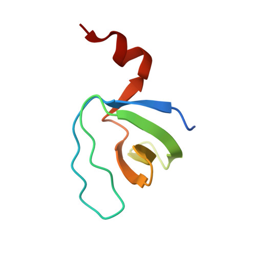
    4ESR

  • PubMed Abstract: 

    ABL tyrosine kinase inhibitor (TKI) therapy induces clinical remission in chronic myeloid leukemia (CML) patients but early relapses and later emergence of TKI-resistant disease remain problematic. We recently demonstrated that the AHI-1 oncogene physically interacts with BCR-ABL and JAK2 and mediates cellular resistance to TKI in CML stem/progenitor cells. We now show that deletion of the SH3 domain of AHI-1 significantly enhances apoptotic response of BCR-ABL(+) cells to TKIs compared to cells expressing full-length AHI-1. We have also discovered a novel interaction between AHI-1 and Dynamin-2, a GTPase, through the AHI-1 SH3 domain. The crystal structure of the AHI-1 SH3 domain at 1.53-Å resolution reveals that it adopts canonical SH3 folding, with the exception of an unusual C-terminal α helix. PD1R peptide, known to interact with the PI3K SH3 domain, was used to model the binding pattern between the AHI-1 SH3 domain and its ligands. These studies showed that an "Arg-Arg-Trp" stack may form within the binding interface, providing a potential target site for designing specific drugs. The crystal structure of the AHI-1 SH3 domain thus provides a valuable tool for identification of key interaction sites in regulation of drug resistance and for the development of small molecule inhibitors for CML.


  • Organizational Affiliation

    Terry Fox Laboratory, British Columbia Cancer Agency, Vancouver, BC, Canada.


Macromolecules
Find similar proteins by:  (by identity cutoff)  |  3D Structure
Entity ID: 1
MoleculeChains Sequence LengthOrganismDetailsImage
Jouberin
A, B
69Homo sapiensMutation(s): 0 
Gene Names: AHI1
UniProt & NIH Common Fund Data Resources
Find proteins for Q8N157 (Homo sapiens)
Explore Q8N157 
Go to UniProtKB:  Q8N157
PHAROS:  Q8N157
GTEx:  ENSG00000135541 
Entity Groups  
Sequence Clusters30% Identity50% Identity70% Identity90% Identity95% Identity100% Identity
UniProt GroupQ8N157
Sequence Annotations
Expand
  • Reference Sequence
Experimental Data & Validation

Experimental Data

  • Method: X-RAY DIFFRACTION
  • Resolution: 1.53 Å
  • R-Value Free: 0.139 
  • R-Value Work: 0.124 
  • R-Value Observed: 0.125 
  • Space Group: P 41 21 2
Unit Cell:
Length ( Å )Angle ( ˚ )
a = 68.142α = 90
b = 68.142β = 90
c = 98.489γ = 90
Software Package:
Software NamePurpose
DENZOdata reduction
SCALEPACKdata scaling
REFMACrefinement
PDB_EXTRACTdata extraction
HKL-2000data collection
HKL-2000data reduction
PHASERphasing

Structure Validation

View Full Validation Report


Entry History 

Deposition Data

Revision History  (Full details and data files)

  • Version 1.0: 2012-06-06
    Type: Initial release
  • Version 1.1: 2012-11-14
    Changes: Database references
  • Version 1.2: 2017-11-15
    Changes: Refinement description
  • Version 1.3: 2024-02-28
    Changes: Data collection, Database references, Derived calculations