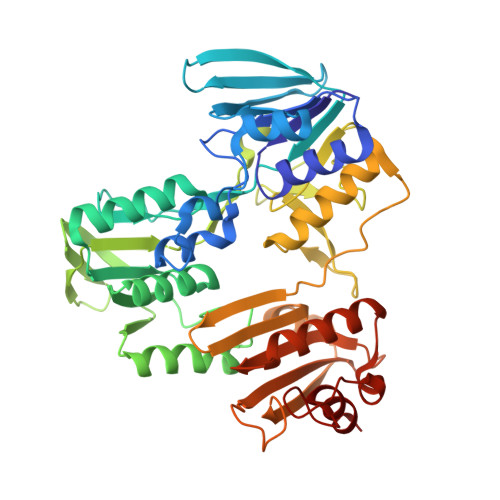
Turnover-Dependent Covalent Inactivation of Staphylococcus aureus Coenzyme A-Disulfide Reductase by Coenzyme A-Mimetics: Mechanistic and Structural Insights.
Wallace, B.D., Edwards, J.S., Wallen, J.R., Moolman, W.J., van der Westhuyzen, R., Strauss, E., Redinbo, M.R., Claiborne, A.(2012) Biochemistry 51: 7699-7711
- PubMed: 22954034
- DOI: https://doi.org/10.1021/bi301026c
- Primary Citation of Related Structures:
4EM3, 4EM4, 4EMW, 4EQR, 4EQS, 4EQW, 4EQX - PubMed Abstract:
Disruption of the unusual thiol-based redox homeostasis mechanisms in Staphylococcus aureus represents a unique opportunity to identify new metabolic processes and new targets for intervention. Targeting uncommon aspects of CoASH biosynthetic and redox functions in S. aureus, the antibiotic CJ-15,801 has recently been demonstrated to be an antimetabolite of the CoASH biosynthetic pathway in this organism; CoAS-mimetics containing α,β-unsaturated sulfone and carboxyl moieties have also been exploited as irreversible inhibitors of S. aureus coenzyme A-disulfide reductase (SaCoADR). In this work we have determined the crystal structures of three of these covalent SaCoADR-inhibitor complexes, prepared by inactivation of wild-type enzyme during turnover. The structures reveal the covalent linkage between the active-site Cys43-S(γ) and C(β) of the vinyl sulfone or carboxyl moiety. The full occupancy of two inhibitor molecules per enzyme dimer, together with kinetic analyses of the wild-type/C43S heterodimer, indicates that half-sites-reactivity is not a factor during normal catalytic turnover. Further, we provide the structures of SaCoADR active-site mutants; in particular, Tyr419'-OH plays dramatic roles in directing intramolecular reduction of the Cys43-SSCoA redox center, in the redox asymmetry observed for the two FAD per dimer in NADPH titrations, and in catalysis. The two conformations observed for the Ser43 side chain in the C43S mutant structure lend support to a conformational switch for Cys43-S(γ) during its catalytic Cys43-SSCoA/Cys43-SH redox cycle. Finally, the structures of the three inhibitor complexes provide a framework for design of more effective inhibitors with therapeutic potential against several major bacterial pathogens.
Organizational Affiliation:
Departments of Chemistry and Biochemistry and Biophysics, University of North Carolina at Chapel Hill, Chapel Hill, North Carolina 27599-3290, United States.


