4EQ7

Structure of Atu4243-GABA receptor


Experimental Data Snapshot

  • Method: X-RAY DIFFRACTION
  • Resolution: 1.91 Å
  • R-Value Free: 0.192 
  • R-Value Work: 0.166 
  • R-Value Observed: 0.167 

wwPDB Validation   3D Report Full Report


This is version 1.1 of the entry. See complete history


Literature

Structural basis for selective GABA binding in bacterial pathogens.

Planamente, S.Mondy, S.Hommais, F.Vigouroux, A.Morera, S.Faure, D.

(2012) Mol Microbiol 86: 1085-1099

  • DOI: https://doi.org/10.1111/mmi.12043
  • Primary Citation of Related Structures:  
    4EQ7, 4EUO

  • PubMed Abstract: 

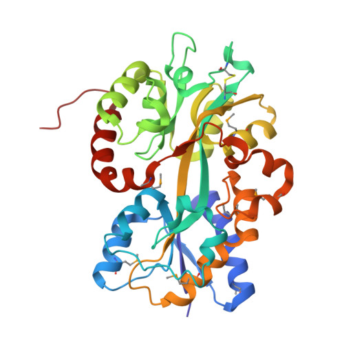
    GABA acts as an intercellular signal in eukaryotes and as an interspecies signal in host-microbe interactions. Structural characteristics of selective eukaryotic GABA receptors and bacterial GABA sensors are unknown. Here, we identified the selective GABA-binding protein, called Atu4243, in the plant pathogen Agrobacterium tumefaciens. A constructed atu4243 mutant was affected in GABA transport and in expression of the GABA-regulated functions, including aggressiveness on two plant hosts and degradation of the quorum-sensing signal. The GABA-bound Atu4243 structure at 1.28 Å reveals that GABA adopts a conformation never observed so far and interacts with two key residues, Arg(203) and Asp(226) of which the role in GABA binding and GABA signalling in Agrobacterium has been validated using appropriate mutants. The conformational GABA-analogue trans-4-aminocrotonic acid (TACA) antagonizes GABA activity, suggesting structural similarities between the binding sites of the bacterial sensor Atu4243 and mammalian GABA(C) receptors. Exploration of genomic databases reveals Atu4243 orthologues in several pathogenic and symbiotic proteobacteria, such as Rhizobium, Azospirillum, Burkholderia and Pseudomonas. Thus, this study establishes a structural basis for selective GABA sensors and offers opportunities for deciphering the role of the GABA-mediated communication in several host-pathogen interactions.


  • Organizational Affiliation

    Institut des Sciences du Végétal, CNRS, avenue de la terrasse, 91198, Gif-sur-Yvette, France.


Macromolecules
Find similar proteins by:  (by identity cutoff)  |  3D Structure
Entity ID: 1
MoleculeChains Sequence LengthOrganismDetailsImage
ABC transporter, substrate binding protein (Polyamine)
A, B
320Agrobacterium fabrum str. C58Mutation(s): 0 
Gene Names: Atu4243
UniProt
Find proteins for A9CGA5 (Agrobacterium fabrum (strain C58 / ATCC 33970))
Explore A9CGA5 
Go to UniProtKB:  A9CGA5
Entity Groups  
Sequence Clusters30% Identity50% Identity70% Identity90% Identity95% Identity100% Identity
UniProt GroupA9CGA5
Sequence Annotations
Expand
  • Reference Sequence
Small Molecules
Ligands 2 Unique
IDChains Name / Formula / InChI Key2D Diagram3D Interactions
SO4
Query on SO4

Download Ideal Coordinates CCD File 
C [auth A]
D [auth A]
E [auth A]
F [auth A]
G [auth A]
C [auth A],
D [auth A],
E [auth A],
F [auth A],
G [auth A],
J [auth B],
K [auth B],
L [auth B],
M [auth B]
SULFATE ION
O4 S
QAOWNCQODCNURD-UHFFFAOYSA-L
GOL
Query on GOL

Download Ideal Coordinates CCD File 
H [auth A],
I [auth A],
N [auth B],
O [auth B],
P [auth B]
GLYCEROL
C3 H8 O3
PEDCQBHIVMGVHV-UHFFFAOYSA-N
Modified Residues  1 Unique
IDChains TypeFormula2D DiagramParent
MSE
Query on MSE
A, B
L-PEPTIDE LINKINGC5 H11 N O2 SeMET
Experimental Data & Validation

Experimental Data

  • Method: X-RAY DIFFRACTION
  • Resolution: 1.91 Å
  • R-Value Free: 0.192 
  • R-Value Work: 0.166 
  • R-Value Observed: 0.167 
  • Space Group: P 61 2 2
Unit Cell:
Length ( Å )Angle ( ˚ )
a = 104.28α = 90
b = 104.28β = 90
c = 286.71γ = 120
Software Package:
Software NamePurpose
DNAdata collection
SHELXSphasing
BUSTERrefinement
XDSdata reduction
XDSdata scaling

Structure Validation

View Full Validation Report


Entry History 

Deposition Data

Revision History  (Full details and data files)

  • Version 1.0: 2012-11-21
    Type: Initial release
  • Version 1.1: 2012-12-19
    Changes: Database references