4EMJ

Complex between the reductase and ferredoxin components of toluene dioxygenase


Experimental Data Snapshot

  • Method: X-RAY DIFFRACTION
  • Resolution: 2.40 Å
  • R-Value Free: 0.234 
  • R-Value Work: 0.169 
  • R-Value Observed: 0.172 

wwPDB Validation   3D Report Full Report


Ligand Structure Quality Assessment 


This is version 1.3 of the entry. See complete history


Literature

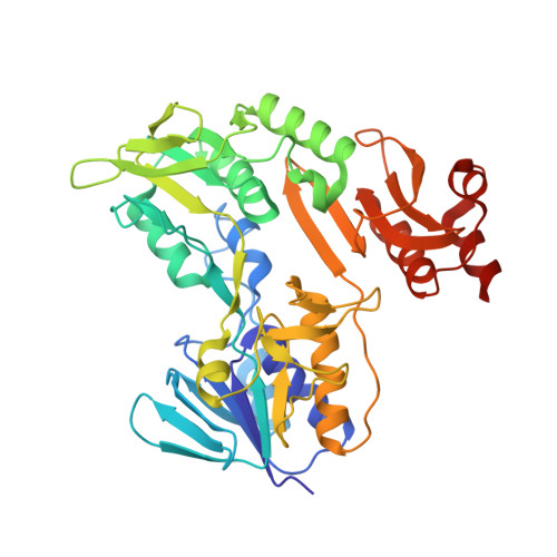
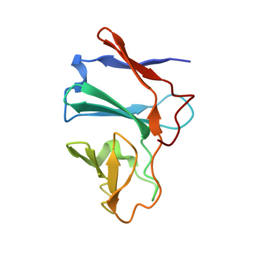
Suppression of Electron Transfer to Dioxygen by Charge Transfer and Electron Transfer Complexes in the FAD-dependent Reductase Component of Toluene Dioxygenase.

Lin, T.Y.Werther, T.Jeoung, J.H.Dobbek, H.

(2012) J Biol Chem 287: 38338-38346

  • DOI: https://doi.org/10.1074/jbc.M112.374918
  • Primary Citation of Related Structures:  
    4EMI, 4EMJ

  • PubMed Abstract: 

    The three-component toluene dioxygenase system consists of an FAD-containing reductase, a Rieske-type [2Fe-2S] ferredoxin, and a Rieske-type dioxygenase. The task of the FAD-containing reductase is to shuttle electrons from NADH to the ferredoxin, a reaction the enzyme has to catalyze in the presence of dioxygen. We investigated the kinetics of the reductase in the reductive and oxidative half-reaction and detected a stable charge transfer complex between the reduced reductase and NAD(+) at the end of the reductive half-reaction, which is substantially less reactive toward dioxygen than the reduced reductase in the absence of NAD(+). A plausible reason for the low reactivity toward dioxygen is revealed by the crystal structure of the complex between NAD(+) and reduced reductase, which shows that the nicotinamide ring and the protein matrix shield the reactive C4a position of the isoalloxazine ring and force the tricycle into an atypical planar conformation, both factors disfavoring the reaction of the reduced flavin with dioxygen. A rapid electron transfer from the charge transfer complex to electron acceptors further reduces the risk of unwanted side reactions, and the crystal structure of a complex between the reductase and its cognate ferredoxin shows a short distance between the electron-donating and -accepting cofactors. Attraction between the two proteins is likely mediated by opposite charges at one large patch of the complex interface. The stability, specificity, and reactivity of the observed charge transfer and electron transfer complexes are thought to prevent the reaction of reductase(TOL) with dioxygen and thus present a solution toward conflicting requirements.


  • Organizational Affiliation

    Institut für Biologie, Strukturbiologie/Biochemie, Humboldt-Universität zu Berlin, D-10115 Berlin, Germany.


Macromolecules
Find similar proteins by:  (by identity cutoff)  |  3D Structure
Entity ID: 1
MoleculeChains Sequence LengthOrganismDetailsImage
TodA410Pseudomonas putidaMutation(s): 0 
Gene Names: tobAtodA
UniProt
Find proteins for A5W4E9 (Pseudomonas putida (strain ATCC 700007 / DSM 6899 / BCRC 17059 / F1))
Explore A5W4E9 
Go to UniProtKB:  A5W4E9
Entity Groups  
Sequence Clusters30% Identity50% Identity70% Identity90% Identity95% Identity100% Identity
UniProt GroupA5W4E9
Sequence Annotations
Expand
  • Reference Sequence
Find similar proteins by:  (by identity cutoff)  |  3D Structure
Entity ID: 2
MoleculeChains Sequence LengthOrganismDetailsImage
Toluene 1,2-dioxygenase system ferredoxin subunit107Pseudomonas putidaMutation(s): 0 
Gene Names: todB
UniProt
Find proteins for A5W4F0 (Pseudomonas putida (strain ATCC 700007 / DSM 6899 / BCRC 17059 / F1))
Explore A5W4F0 
Go to UniProtKB:  A5W4F0
Entity Groups  
Sequence Clusters30% Identity50% Identity70% Identity90% Identity95% Identity100% Identity
UniProt GroupA5W4F0
Sequence Annotations
Expand
  • Reference Sequence
Small Molecules
Ligands 2 Unique
IDChains Name / Formula / InChI Key2D Diagram3D Interactions
FAD
Query on FAD

Download Ideal Coordinates CCD File 
C [auth A]FLAVIN-ADENINE DINUCLEOTIDE
C27 H33 N9 O15 P2
VWWQXMAJTJZDQX-UYBVJOGSSA-N
FES
Query on FES

Download Ideal Coordinates CCD File 
D [auth B]FE2/S2 (INORGANIC) CLUSTER
Fe2 S2
NIXDOXVAJZFRNF-UHFFFAOYSA-N
Experimental Data & Validation

Experimental Data

  • Method: X-RAY DIFFRACTION
  • Resolution: 2.40 Å
  • R-Value Free: 0.234 
  • R-Value Work: 0.169 
  • R-Value Observed: 0.172 
  • Space Group: P 65
Unit Cell:
Length ( Å )Angle ( ˚ )
a = 120.29α = 90
b = 120.29β = 90
c = 60.42γ = 120
Software Package:
Software NamePurpose
MAR345data collection
PHASERphasing
PHENIXrefinement
XDSdata reduction
XSCALEdata scaling

Structure Validation

View Full Validation Report


Ligand Structure Quality Assessment 


Entry History 

Deposition Data

Revision History  (Full details and data files)

  • Version 1.0: 2012-09-26
    Type: Initial release
  • Version 1.1: 2012-11-21
    Changes: Database references
  • Version 1.2: 2017-11-15
    Changes: Refinement description
  • Version 1.3: 2024-02-28
    Changes: Data collection, Database references, Derived calculations