4EHW

Crystal structure of LpxK from Aquifex aeolicus at 2.3 angstrom resolution


Experimental Data Snapshot

  • Method: X-RAY DIFFRACTION
  • Resolution: 2.30 Å
  • R-Value Free: 0.263 
  • R-Value Work: 0.228 
  • R-Value Observed: 0.232 

wwPDB Validation   3D Report Full Report


This is version 1.1 of the entry. See complete history


Literature

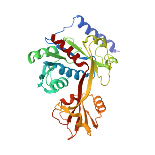
Crystal structure of LpxK, the 4'-kinase of lipid A biosynthesis and atypical P-loop kinase functioning at the membrane interface.

Emptage, R.P.Daughtry, K.D.Pemble, C.W.Raetz, C.R.

(2012) Proc Natl Acad Sci U S A 109: 12956-12961

  • DOI: https://doi.org/10.1073/pnas.1206072109
  • Primary Citation of Related Structures:  
    4EHW, 4EHX, 4EHY

  • PubMed Abstract: 

    In Gram-negative bacteria, the hydrophobic anchor of the outer membrane lipopolysaccharide is lipid A, a saccharolipid that plays key roles in both viability and pathogenicity of these organisms. The tetraacyldisaccharide 4'-kinase (LpxK) of the diverse P-loop-containing nucleoside triphosphate hydrolase superfamily catalyzes the sixth step in the biosynthetic pathway of lipid A, and is the only known P-loop kinase to act upon a lipid substrate at the membrane. Here, we report the crystal structures of apo- and ADP/Mg(2+)-bound forms of Aquifex aeolicus LpxK to a resolution of 1.9 Å and 2.2 Å, respectively. LpxK consists of two α/β/α sandwich domains connected by a two-stranded β-sheet linker. The N-terminal domain, which has most structural homology to other family members, is responsible for catalysis at the P-loop and positioning of the disaccharide-1-phosphate substrate for phosphoryl transfer on the inner membrane. The smaller C-terminal domain, a substructure unique to LpxK, helps bind the nucleotide substrate and Mg(2+) cation using a 25° hinge motion about its base. Activity was severely reduced in alanine point mutants of conserved residues D138 and D139, which are not directly involved in ADP or Mg(2+) binding in our structures, indicating possible roles in phosphoryl acceptor positioning or catalysis. Combined structural and kinetic studies have led to an increased understanding of the enzymatic mechanism of LpxK and provided the framework for structure-based antimicrobial design.


  • Organizational Affiliation

    Department of Biochemistry and Human Vaccine Institute, Duke University Medical Center, Durham, NC 27710, USA.


Macromolecules
Find similar proteins by:  (by identity cutoff)  |  3D Structure
Entity ID: 1
MoleculeChains Sequence LengthOrganismDetailsImage
Tetraacyldisaccharide 4'-kinase317Aquifex aeolicus VF5Mutation(s): 0 
Gene Names: aq_1656lpxK
EC: 2.7.1.130
UniProt
Find proteins for O67572 (Aquifex aeolicus (strain VF5))
Explore O67572 
Go to UniProtKB:  O67572
Entity Groups  
Sequence Clusters30% Identity50% Identity70% Identity90% Identity95% Identity100% Identity
UniProt GroupO67572
Sequence Annotations
Expand
  • Reference Sequence
Small Molecules
Ligands 2 Unique
IDChains Name / Formula / InChI Key2D Diagram3D Interactions
MPD
Query on MPD

Download Ideal Coordinates CCD File 
D [auth A],
E [auth A],
G [auth A],
J [auth A]
(4S)-2-METHYL-2,4-PENTANEDIOL
C6 H14 O2
SVTBMSDMJJWYQN-YFKPBYRVSA-N
GOL
Query on GOL

Download Ideal Coordinates CCD File 
B [auth A]
C [auth A]
F [auth A]
H [auth A]
I [auth A]
B [auth A],
C [auth A],
F [auth A],
H [auth A],
I [auth A],
K [auth A]
GLYCEROL
C3 H8 O3
PEDCQBHIVMGVHV-UHFFFAOYSA-N
Experimental Data & Validation

Experimental Data

  • Method: X-RAY DIFFRACTION
  • Resolution: 2.30 Å
  • R-Value Free: 0.263 
  • R-Value Work: 0.228 
  • R-Value Observed: 0.232 
  • Space Group: P 41 21 2
Unit Cell:
Length ( Å )Angle ( ˚ )
a = 101.191α = 90
b = 101.191β = 90
c = 131.904γ = 90
Software Package:
Software NamePurpose
d*TREKdata reduction
PHENIXrefinement
PDB_EXTRACTdata extraction
StructureStudiodata collection
d*TREKdata scaling
SOLVEphasing

Structure Validation

View Full Validation Report


Entry History 

Deposition Data

Revision History  (Full details and data files)

  • Version 1.0: 2012-08-29
    Type: Initial release
  • Version 1.1: 2024-02-28
    Changes: Data collection, Database references, Derived calculations