


Crystal structure of the Ubl domain of BAG6
Kozlov, G., LePage, K., Vinaik, R., Gehring, K.To be published.
Experimental Data Snapshot
wwPDB Validation 3D Report Full Report

Entity ID: 1 | |||||
|---|---|---|---|---|---|
| Molecule | Chains | Sequence Length | Organism | Details | Image |
| Large proline-rich protein BAG6 | 88 | Homo sapiens | Mutation(s): 0 Gene Names: BAG6, BAT3, G3 | 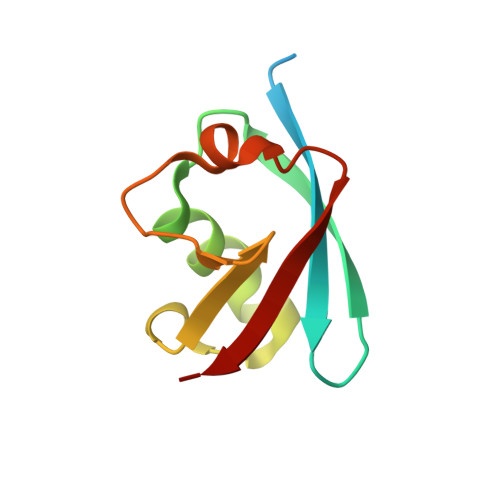 | |
UniProt & NIH Common Fund Data Resources | |||||
Find proteins for P46379 (Homo sapiens) Explore P46379 Go to UniProtKB: P46379 | |||||
PHAROS: P46379 GTEx: ENSG00000204463 | |||||
Entity Groups | |||||
| Sequence Clusters | 30% Identity50% Identity70% Identity90% Identity95% Identity100% Identity | ||||
| UniProt Group | P46379 | ||||
Sequence AnnotationsExpand | |||||
| |||||
| Length ( Å ) | Angle ( ˚ ) |
|---|---|
| a = 30.572 | α = 90 |
| b = 43.193 | β = 104.33 |
| c = 51.734 | γ = 90 |
| Software Name | Purpose |
|---|---|
| ADSC | data collection |
| PHASER | phasing |
| REFMAC | refinement |
| HKL-2000 | data reduction |
| HKL-2000 | data scaling |
RCSB PDB (citation) is hosted by
RCSB PDB is a member of the

