4EAY

Crystal structures of mannonate dehydratase from Escherichia coli strain K12 complexed with D-mannonate


Experimental Data Snapshot

  • Method: X-RAY DIFFRACTION
  • Resolution: 2.35 Å
  • R-Value Free: 0.236 
  • R-Value Work: 0.189 
  • R-Value Observed: 0.191 

wwPDB Validation   3D Report Full Report


Ligand Structure Quality Assessment 


This is version 1.1 of the entry. See complete history


Literature

Structural insights into decreased enzymatic activity induced by an insert sequence in mannonate dehydratase from Gram negative bacterium.

Qiu, X.Tao, Y.Zhu, Y.Yuan, Y.Zhang, Y.Liu, H.Gao, Y.Teng, M.Niu, L.

(2012) J Struct Biol 180: 327-334

  • DOI: https://doi.org/10.1016/j.jsb.2012.06.013
  • Primary Citation of Related Structures:  
    4EAC, 4EAY

  • PubMed Abstract: 

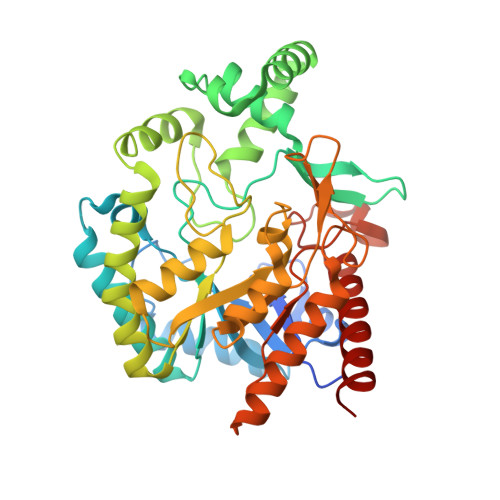
    Mannonate dehydratase (ManD; EC4.2.1.8) catalyzes the dehydration of D-mannonate to 2-keto-3-deoxygluconate. It is the third enzyme in the pathway for dissimilation of D-glucuronate to 2-keto-3-deoxygluconate involving in the Entner-Doudoroff pathway in certain bacterial and archaeal species. ManD from Gram negative bacteria has an insert sequence as compared to those from Gram positives revealed by sequence analysis. To evaluate the impact of this insert sequence on the catalytic efficiency, we solved the crystal structures of ManD from Escherichia coli strain K12 and its complex with D-mannonate, which reveal that this insert sequence forms two α helices locating above the active site. The two insert α helices introduce a loop that forms a cap covering the substrate binding pocket, which restricts the tunnels of substrate entering and product releasing from the active site. Site-directed mutations and enzymatic activity assays confirm that the catalytic rate is decreased by this loop. These features are conserved among Gram negative bacteria. Thus, the insert sequence of ManD from Gram negative bacteria acts as a common inducer to decrease the catalytic rate and consequently the glucuronate metabolic rate as compared to those from Gram positives. Moreover, residues essential for substrate to enter the active site were characterized via structural analysis and enzymatic activity assays.


  • Organizational Affiliation

    Hefei National Laboratory for Physical Sciences at Microscale and School of Life Sciences, University of Science and Technology of China, Key Laboratory of Structural Biology, Chinese Academy of Sciences, Hefei, Anhui, PR China.


Macromolecules
Find similar proteins by:  (by identity cutoff)  |  3D Structure
Entity ID: 1
MoleculeChains Sequence LengthOrganismDetailsImage
Mannonate dehydratase
A, B, C, D
414Escherichia coli K-12Mutation(s): 0 
Gene Names: uxuAb4322JW4285
EC: 4.2.1.8
UniProt
Find proteins for P24215 (Escherichia coli (strain K12))
Explore P24215 
Go to UniProtKB:  P24215
Entity Groups  
Sequence Clusters30% Identity50% Identity70% Identity90% Identity95% Identity100% Identity
UniProt GroupP24215
Sequence Annotations
Expand
  • Reference Sequence
Small Molecules
Ligands 3 Unique
IDChains Name / Formula / InChI Key2D Diagram3D Interactions
CS2
Query on CS2

Download Ideal Coordinates CCD File 
F [auth A],
I [auth B]
D-MANNONIC ACID
C6 H12 O7
RGHNJXZEOKUKBD-MBMOQRBOSA-N
MN
Query on MN

Download Ideal Coordinates CCD File 
G [auth A],
J [auth B],
L [auth C],
N [auth D]
MANGANESE (II) ION
Mn
WAEMQWOKJMHJLA-UHFFFAOYSA-N
CL
Query on CL

Download Ideal Coordinates CCD File 
E [auth A],
H [auth B],
K [auth C],
M [auth D]
CHLORIDE ION
Cl
VEXZGXHMUGYJMC-UHFFFAOYSA-M
Experimental Data & Validation

Experimental Data

  • Method: X-RAY DIFFRACTION
  • Resolution: 2.35 Å
  • R-Value Free: 0.236 
  • R-Value Work: 0.189 
  • R-Value Observed: 0.191 
  • Space Group: P 21 21 2
Unit Cell:
Length ( Å )Angle ( ˚ )
a = 159.47α = 90
b = 238.58β = 90
c = 54.47γ = 90
Software Package:
Software NamePurpose
ADSCdata collection
PHASERphasing
REFMACrefinement
iMOSFLMdata reduction
SCALAdata scaling

Structure Validation

View Full Validation Report


Ligand Structure Quality Assessment 


Entry History 

Deposition Data

Revision History  (Full details and data files)

  • Version 1.0: 2013-03-27
    Type: Initial release
  • Version 1.1: 2024-03-20
    Changes: Data collection, Database references, Derived calculations, Refinement description