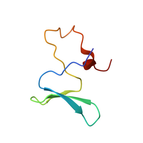
Dimerization and heme binding are conserved in amphibian and starfish homologues of the microRNA processing protein DGCR8.
Senturia, R., Laganowsky, A., Barr, I., Scheidemantle, B.D., Guo, F.(2012) PLoS One 7: e39688-e39688
- PubMed: 22768307
- DOI: https://doi.org/10.1371/journal.pone.0039688
- Primary Citation of Related Structures:
4E5R - PubMed Abstract:
Human DiGeorge Critical Region 8 (DGCR8) is an essential microRNA (miRNA) processing factor that is activated via direct interaction with Fe(III) heme. In order for DGCR8 to bind heme, it must dimerize using a dimerization domain embedded within its heme-binding domain (HBD). We previously reported a crystal structure of the dimerization domain from human DGCR8, which demonstrated how dimerization results in the formation of a surface important for association with heme. Here, in an attempt to crystallize the HBD, we search for DGCR8 homologues and show that DGCR8 from Patiria miniata (bat star) also binds heme. The extinction coefficients (ε) of DGCR8-heme complexes are determined; these values are useful for biochemical analyses and allow us to estimate the heme occupancy of DGCR8 proteins. Additionally, we present the crystal structure of the Xenopus laevis dimerization domain. The structure is very similar to that of human DGCR8. Our results indicate that dimerization and heme binding are evolutionarily conserved properties of DGCR8 homologues not only in vertebrates, but also in at least some invertebrates.
Organizational Affiliation:
Department of Biological Chemistry, David Geffen School of Medicine, Molecular Biology Institute, University of California Los Angeles, Los Angeles, California, USA.