4DRS

Crystal structure of Cryptosporidium parvum pyruvate kinase


Experimental Data Snapshot

  • Method: X-RAY DIFFRACTION
  • Resolution: 2.50 Å
  • R-Value Free: 0.249 
  • R-Value Work: 0.213 
  • R-Value Observed: 0.215 

wwPDB Validation   3D Report Full Report


This is version 1.3 of the entry. See complete history


Literature

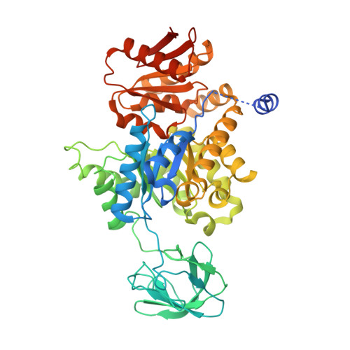
Crystal structure of Cryptosporidium parvum pyruvate kinase.

Cook, W.J.Senkovich, O.Aleem, K.Chattopadhyay, D.

(2012) PLoS One 7: e46875-e46875

  • DOI: https://doi.org/10.1371/journal.pone.0046875
  • Primary Citation of Related Structures:  
    4DRS

  • PubMed Abstract: 

    Pyruvate kinase plays a critical role in cellular metabolism of glucose by serving as a major regulator of glycolysis. This tetrameric enzyme is allosterically regulated by different effector molecules, mainly phosphosugars. In response to binding of effector molecules and substrates, significant structural changes have been identified in various pyruvate kinase structures. Pyruvate kinase of Cryptosporidium parvum is exceptional among known enzymes of protozoan origin in that it exhibits no allosteric property in the presence of commonly known effector molecules. The crystal structure of pyruvate kinase from C. parvum has been solved by molecular replacement techniques and refined to 2.5 Å resolution. In the active site a glycerol molecule is located near the γ-phosphate site of ATP, and the protein structure displays a partially closed active site. However, unlike other structures where the active site is closed, the α6' helix in C. parvum pyruvate kinase unwinds and assumes an extended conformation. In the crystal structure a sulfate ion is found at a site that is occupied by a phosphate of the effector molecule in many pyruvate kinase structures. A new feature of the C. parvum pyruvate kinase structure is the presence of a disulfide bond cross-linking the two monomers in the asymmetric unit. The disulfide bond is formed between cysteine residue 26 in the short N-helix of one monomer with cysteine residue 312 in a long helix (residues 303-320) of the second monomer at the interface of these monomers. Both cysteine residues are unique to C. parvum, and the disulfide bond remained intact in a reduced environment. However, the significance of this bond, if any, remains unknown at this time.


  • Organizational Affiliation

    Department of Pathology, University of Alabama at Birmingham, Birmingham, Alabama, United States of America.


Macromolecules
Find similar proteins by:  (by identity cutoff)  |  3D Structure
Entity ID: 1
MoleculeChains Sequence LengthOrganismDetailsImage
Pyruvate kinase
A, B
526Cryptosporidium parvum Iowa IIMutation(s): 0 
Gene Names: cgd1_2040
EC: 2.7.1.40
UniProt
Find proteins for Q5CSM7 (Cryptosporidium parvum (strain Iowa II))
Explore Q5CSM7 
Go to UniProtKB:  Q5CSM7
Entity Groups  
Sequence Clusters30% Identity50% Identity70% Identity90% Identity95% Identity100% Identity
UniProt GroupQ5CSM7
Sequence Annotations
Expand
  • Reference Sequence
Small Molecules
Ligands 3 Unique
IDChains Name / Formula / InChI Key2D Diagram3D Interactions
SO4
Query on SO4

Download Ideal Coordinates CCD File 
C [auth A],
D [auth A],
J [auth B],
K [auth B]
SULFATE ION
O4 S
QAOWNCQODCNURD-UHFFFAOYSA-L
GOL
Query on GOL

Download Ideal Coordinates CCD File 
E [auth A],
F [auth A],
L [auth B],
M [auth B]
GLYCEROL
C3 H8 O3
PEDCQBHIVMGVHV-UHFFFAOYSA-N
ACT
Query on ACT

Download Ideal Coordinates CCD File 
G [auth A]
H [auth A]
I [auth A]
N [auth B]
O [auth B]
G [auth A],
H [auth A],
I [auth A],
N [auth B],
O [auth B],
P [auth B]
ACETATE ION
C2 H3 O2
QTBSBXVTEAMEQO-UHFFFAOYSA-M
Experimental Data & Validation

Experimental Data

  • Method: X-RAY DIFFRACTION
  • Resolution: 2.50 Å
  • R-Value Free: 0.249 
  • R-Value Work: 0.213 
  • R-Value Observed: 0.215 
  • Space Group: P 21 21 2
Unit Cell:
Length ( Å )Angle ( ˚ )
a = 129.895α = 90
b = 136.941β = 90
c = 77.233γ = 90
Software Package:
Software NamePurpose
REFMACrefinement
CNSrefinement
HKL-2000data collection
HKL-2000data reduction
HKL-2000data scaling
CNSphasing

Structure Validation

View Full Validation Report


Entry History 

Deposition Data

Revision History  (Full details and data files)

  • Version 1.0: 2012-10-17
    Type: Initial release
  • Version 1.1: 2013-01-23
    Changes: Database references
  • Version 1.2: 2019-07-17
    Changes: Data collection, Refinement description
  • Version 1.3: 2023-09-13
    Changes: Data collection, Database references, Derived calculations, Refinement description