4DL1

Crystal Structure of human Myeloperoxidase with covalent thioxanthine analog


Experimental Data Snapshot

  • Method: X-RAY DIFFRACTION
  • Resolution: 2.00 Å
  • R-Value Free: 0.246 
  • R-Value Work: 0.190 
  • R-Value Observed: 0.193 

wwPDB Validation   3D Report Full Report


Ligand Structure Quality Assessment 


This is version 2.0 of the entry. See complete history


Literature

Deconstruction of activity-dependent covalent modification of heme in human neutrophil myeloperoxidase by multistage mass spectrometry (MS(4)).

Geoghegan, K.F.Varghese, A.H.Feng, X.Bessire, A.J.Conboy, J.J.Ruggeri, R.B.Ahn, K.Spath, S.N.Filippov, S.V.Conrad, S.J.Carpino, P.A.Guimaraes, C.R.Vajdos, F.F.

(2012) Biochemistry 51: 2065-2077

  • DOI: https://doi.org/10.1021/bi201872j
  • Primary Citation of Related Structures:  
    4DL1

  • PubMed Abstract: 

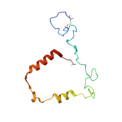
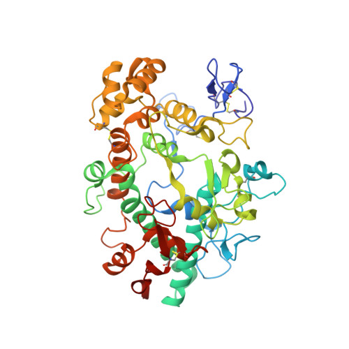
    Myeloperoxidase (MPO) is known to be inactivated and covalently modified by treatment with hydrogen peroxide and agents similar to 3-(2-ethoxypropyl)-2-thioxo-2,3-dihydro-1H-purin-6(9H)-one (1), a 254.08 Da derivative of 2-thioxanthine. Peptide mapping by liquid chromatography and mass spectrometry detected modification by 1 in a labile peptide-heme-peptide fragment of the enzyme, accompanied by a mass increase of 252.08 Da. The loss of two hydrogen atoms was consistent with mechanism-based oxidative coupling. Multistage mass spectrometry (MS(4)) of the modified fragment in an ion trap/Orbitrap spectrometer demonstrated that 1 was coupled directly to heme. Use of a 10 amu window delivered the full isotopic envelope of each precursor ion to collision-induced dissociation, preserving definitive isotopic profiles for iron-containing fragments through successive steps of multistage mass spectrometry. Iron isotope signatures and accurate mass measurements supported the structural assignments. Crystallographic analysis confirmed linkage between the methyl substituent of the heme pyrrole D ring and the sulfur atom of 1. The final orientation of 1 perpendicular to the plane of the heme ring suggested a mechanism consisting of two consecutive one-electron oxidations of 1 by MPO. Multistage mass spectrometry using stage-specific collision energies permits stepwise deconstruction of modifications of heme enzymes containing covalent links between the heme group and the polypeptide chain.


  • Organizational Affiliation

    Pfizer Worldwide Research, Groton, Connecticut 06340, United States. kieran.f.geoghegan@pfizer.com


Macromolecules
Find similar proteins by:  (by identity cutoff)  |  3D Structure
Entity ID: 1
MoleculeChains Sequence LengthOrganismDetailsImage
Myeloperoxidase light chain104Homo sapiensMutation(s): 0 
EC: 1.11.2.2
UniProt & NIH Common Fund Data Resources
Find proteins for P05164 (Homo sapiens)
Explore P05164 
Go to UniProtKB:  P05164
PHAROS:  P05164
GTEx:  ENSG00000005381 
Entity Groups  
Sequence Clusters30% Identity50% Identity70% Identity90% Identity95% Identity100% Identity
UniProt GroupP05164
Sequence Annotations
Expand
  • Reference Sequence
Find similar proteins by:  (by identity cutoff)  |  3D Structure
Entity ID: 2
MoleculeChains Sequence LengthOrganismDetailsImage
Myeloperoxidase heavy chain466Homo sapiensMutation(s): 0 
EC: 1.11.2.2
UniProt & NIH Common Fund Data Resources
Find proteins for P05164 (Homo sapiens)
Explore P05164 
Go to UniProtKB:  P05164
PHAROS:  P05164
GTEx:  ENSG00000005381 
Entity Groups  
Sequence Clusters30% Identity50% Identity70% Identity90% Identity95% Identity100% Identity
UniProt GroupP05164
Sequence Annotations
Expand
  • Reference Sequence
Oligosaccharides

Help

Entity ID: 3
MoleculeChains Length2D Diagram Glycosylation3D Interactions
alpha-D-mannopyranose-(1-3)-[alpha-D-mannopyranose-(1-6)]beta-D-mannopyranose-(1-4)-2-acetamido-2-deoxy-beta-D-glucopyranose-(1-4)-[alpha-L-fucopyranose-(1-6)]2-acetamido-2-deoxy-beta-D-glucopyranose
Q, R, S, T, U
Q, R, S, T, U, V, W, X
6N-Glycosylation
Glycosylation Resources
GlyTouCan:  G82348BZ
GlyCosmos:  G82348BZ
GlyGen:  G82348BZ
Entity ID: 4
MoleculeChains Length2D Diagram Glycosylation3D Interactions
alpha-D-mannopyranose-(1-3)-[alpha-D-mannopyranose-(1-6)]beta-D-mannopyranose-(1-4)-2-acetamido-2-deoxy-beta-D-glucopyranose-(1-4)-2-acetamido-2-deoxy-beta-D-glucopyranose
Y
5N-Glycosylation
Glycosylation Resources
GlyTouCan:  G22768VO
GlyCosmos:  G22768VO
GlyGen:  G22768VO
Small Molecules
Ligands 5 Unique
IDChains Name / Formula / InChI Key2D Diagram3D Interactions
HEM
Query on HEM

Download Ideal Coordinates CCD File 
BA [auth C]
EB [auth J]
GA [auth B]
KB [auth M]
MA [auth E]
BA [auth C],
EB [auth J],
GA [auth B],
KB [auth M],
MA [auth E],
QB [auth N],
SA [auth F],
YA [auth I]
PROTOPORPHYRIN IX CONTAINING FE
C34 H32 Fe N4 O4
KABFMIBPWCXCRK-RGGAHWMASA-L
0KY
Query on 0KY

Download Ideal Coordinates CCD File 
AA [auth A]
FB [auth J]
HA [auth B]
OB [auth O]
QA [auth G]
AA [auth A],
FB [auth J],
HA [auth B],
OB [auth O],
QA [auth G],
TB [auth P],
WA [auth H],
ZA [auth I]
3-[(2R)-2-ethoxypropyl]-2-thioxo-1,2,3,9-tetrahydro-6H-purin-6-one
C10 H14 N4 O2 S
QOTIDWBGIWZLOV-ZCFIWIBFSA-N
NAG
Query on NAG

Download Ideal Coordinates CCD File 
AB [auth K]
BB [auth K]
CA [auth C]
DA [auth C]
GB [auth L]
AB [auth K],
BB [auth K],
CA [auth C],
DA [auth C],
GB [auth L],
HB [auth L],
IA [auth D],
JA [auth D],
LB [auth O],
MB [auth O],
NA [auth G],
OA [auth G],
SB [auth P],
TA [auth H],
UA [auth H]
2-acetamido-2-deoxy-beta-D-glucopyranose
C8 H15 N O6
OVRNDRQMDRJTHS-FMDGEEDCSA-N
CA
Query on CA

Download Ideal Coordinates CCD File 
CB [auth K]
EA [auth C]
IB [auth L]
KA [auth D]
NB [auth O]
CB [auth K],
EA [auth C],
IB [auth L],
KA [auth D],
NB [auth O],
PA [auth G],
RB [auth P],
VA [auth H]
CALCIUM ION
Ca
BHPQYMZQTOCNFJ-UHFFFAOYSA-N
CL
Query on CL

Download Ideal Coordinates CCD File 
DB [auth J]
FA [auth B]
JB [auth M]
LA [auth E]
PB [auth N]
DB [auth J],
FA [auth B],
JB [auth M],
LA [auth E],
PB [auth N],
RA [auth F],
XA [auth I],
Z [auth A]
CHLORIDE ION
Cl
VEXZGXHMUGYJMC-UHFFFAOYSA-M
Modified Residues  1 Unique
IDChains TypeFormula2D DiagramParent
CSO
Query on CSO
B [auth C]
D
F [auth G]
H
J [auth K]
B [auth C],
D,
F [auth G],
H,
J [auth K],
L,
N [auth O],
P
L-PEPTIDE LINKINGC3 H7 N O3 SCYS
Experimental Data & Validation

Experimental Data

  • Method: X-RAY DIFFRACTION
  • Resolution: 2.00 Å
  • R-Value Free: 0.246 
  • R-Value Work: 0.190 
  • R-Value Observed: 0.193 
  • Space Group: P 1 21 1
Unit Cell:
Length ( Å )Angle ( ˚ )
a = 63.829α = 90
b = 242.636β = 91.19
c = 151.505γ = 90
Software Package:
Software NamePurpose
SCALAdata scaling
PHASERphasing
REFMACrefinement
PDB_EXTRACTdata extraction
XSCALEdata scaling

Structure Validation

View Full Validation Report


Ligand Structure Quality Assessment 


Entry History 

Deposition Data

Revision History  (Full details and data files)

  • Version 1.0: 2012-03-21
    Type: Initial release
  • Version 1.1: 2012-05-09
    Changes: Database references
  • Version 2.0: 2020-07-29
    Type: Remediation
    Reason: Carbohydrate remediation
    Changes: Atomic model, Data collection, Derived calculations, Structure summary