4DH2

Crystal structure of Coh-OlpC(Cthe_0452)-Doc435(Cthe_0435) complex: A novel type I Cohesin-Dockerin complex from Clostridium thermocellum ATTC 27405


Experimental Data Snapshot

  • Method: X-RAY DIFFRACTION
  • Resolution: 1.75 Å
  • R-Value Free: 0.215 
  • R-Value Work: 0.181 
  • R-Value Observed: 0.183 

wwPDB Validation   3D Report Full Report


This is version 1.2 of the entry. See complete history


Literature

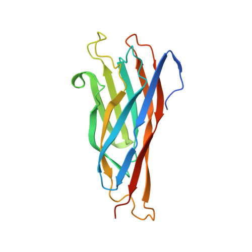
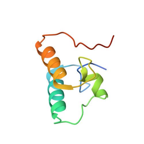
Novel Clostridium thermocellum Type I Cohesin-Dockerin Complexes Reveal a Single Binding Mode.

Bras, J.L.Alves, V.D.Carvalho, A.L.Najmudin, S.Prates, J.A.Ferreira, L.M.Bolam, D.N.Romao, M.J.Gilbert, H.J.Fontes, C.M.

(2012) J Biol Chem 287: 44394-44405

  • DOI: https://doi.org/10.1074/jbc.M112.407700
  • Primary Citation of Related Structures:  
    3UL4, 4DH2

  • PubMed Abstract: 

    Protein-protein interactions play a pivotal role in a large number of biological processes exemplified by the assembly of the cellulosome. Integration of cellulosomal components occurs through the binding of type I cohesin modules located in a non-catalytic molecular scaffold to type I dockerin modules located at the C terminus of cellulosomal enzymes. The majority of type I dockerins display internal symmetry reflected by the presence of two essentially identical cohesin-binding surfaces. Here we report the crystal structures of two novel Clostridium thermocellum type I cohesin-dockerin complexes (CohOlpC-Doc124A and CohOlpA-Doc918). The data revealed that the two dockerins, Doc918 and Doc124A, are unusual because they lack the structural symmetry required to support a dual binding mode. Thus, in both cases, cohesin recognition is dominated by residues located at positions 11, 12, and 19 of one of the dockerin binding surfaces. The alternative binding mode is not possible (Doc918) or highly limited (Doc124A) because residues that assume the critical interacting positions, when dockerins are reoriented by 180°, make steric clashes with the cohesin. In common with a third dockerin (Doc258) that also presents a single binding mode, Doc124A directs the appended cellulase, Cel124A, to the surface of C. thermocellum and not to cellulosomes because it binds preferentially to type I cohesins located at the cell envelope. Although there are a few exceptions, such as Doc918 described here, these data suggest that there is considerable selective pressure for the evolution of a dual binding mode in type I dockerins that direct enzymes into cellulosomes.


  • Organizational Affiliation

    Centro de Investigação Interdisciplinar em Sanidade Animal, Faculdade de Medicina Veterinária, Universidade Técnica de Lisboa, Avenida da Universidade Técnica, 1300-477 Lisboa, Portugal.


Macromolecules
Find similar proteins by:  (by identity cutoff)  |  3D Structure
Entity ID: 1
MoleculeChains Sequence LengthOrganismDetailsImage
Cellulosome anchoring protein cohesin region
A, C
170Acetivibrio thermocellus ATCC 27405Mutation(s): 0 
Gene Names: Cthe_0452
UniProt
Find proteins for A3DCL1 (Acetivibrio thermocellus (strain ATCC 27405 / DSM 1237 / JCM 9322 / NBRC 103400 / NCIMB 10682 / NRRL B-4536 / VPI 7372))
Explore A3DCL1 
Go to UniProtKB:  A3DCL1
Entity Groups  
Sequence Clusters30% Identity50% Identity70% Identity90% Identity95% Identity100% Identity
UniProt GroupA3DCL1
Sequence Annotations
Expand
  • Reference Sequence
Find similar proteins by:  (by identity cutoff)  |  3D Structure
Entity ID: 2
MoleculeChains Sequence LengthOrganismDetailsImage
Dockerin type 1
B, D
82Acetivibrio thermocellus ATCC 27405Mutation(s): 0 
Gene Names: Cthe_0435
UniProt
Find proteins for A3DCJ4 (Acetivibrio thermocellus (strain ATCC 27405 / DSM 1237 / JCM 9322 / NBRC 103400 / NCIMB 10682 / NRRL B-4536 / VPI 7372))
Explore A3DCJ4 
Go to UniProtKB:  A3DCJ4
Entity Groups  
Sequence Clusters30% Identity50% Identity70% Identity90% Identity95% Identity100% Identity
UniProt GroupA3DCJ4
Sequence Annotations
Expand
  • Reference Sequence
Small Molecules
Ligands 2 Unique
IDChains Name / Formula / InChI Key2D Diagram3D Interactions
SO4
Query on SO4

Download Ideal Coordinates CCD File 
E [auth A]
F [auth A]
G [auth A]
J [auth B]
K [auth B]
E [auth A],
F [auth A],
G [auth A],
J [auth B],
K [auth B],
L [auth C],
M [auth C],
N [auth C],
O [auth C]
SULFATE ION
O4 S
QAOWNCQODCNURD-UHFFFAOYSA-L
CA
Query on CA

Download Ideal Coordinates CCD File 
H [auth B],
I [auth B],
P [auth D],
Q [auth D]
CALCIUM ION
Ca
BHPQYMZQTOCNFJ-UHFFFAOYSA-N
Experimental Data & Validation

Experimental Data

  • Method: X-RAY DIFFRACTION
  • Resolution: 1.75 Å
  • R-Value Free: 0.215 
  • R-Value Work: 0.181 
  • R-Value Observed: 0.183 
  • Space Group: P 32 2 1
Unit Cell:
Length ( Å )Angle ( ˚ )
a = 90.76α = 90
b = 90.76β = 90
c = 135.07γ = 120
Software Package:
Software NamePurpose
MOSFLMdata reduction
SCALAdata scaling
DMphasing
PHENIXrefinement
PDB_EXTRACTdata extraction
BALBESphasing

Structure Validation

View Full Validation Report


Entry History 

Deposition Data

Revision History  (Full details and data files)

  • Version 1.0: 2012-11-28
    Type: Initial release
  • Version 1.1: 2013-01-30
    Changes: Database references
  • Version 1.2: 2023-09-13
    Changes: Data collection, Database references, Derived calculations, Refinement description