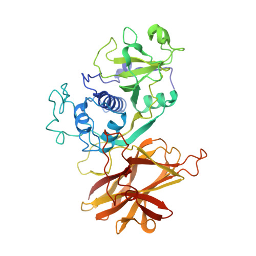
4D5A

Clostridial Cysteine protease Cwp84 C116A after propeptide cleavage


Experimental Data Snapshot

  • Method: X-RAY DIFFRACTION
  • Resolution: 1.60 Å
  • R-Value Free: 0.211 
  • R-Value Work: 0.181 
  • R-Value Observed: 0.183 

wwPDB Validation   3D Report Full Report


This is version 1.3 of the entry. See complete history


Literature

Cwp84, a Clostridium Difficile Cysteine Protease, Exhibits Conformational Flexibility in the Absence of its Propeptide

Bradshaw, W.J.Roberts, A.K.Shone, C.C.Acharya, K.R.

(2015) Acta Crystallogr Sect F Struct Biol Cryst Commun 71: 295

  • DOI: https://doi.org/10.1107/S2053230X15001065
  • Primary Citation of Related Structures:  
    4D59, 4D5A

  • PubMed Abstract: 

    In recent decades, the global healthcare problems caused by Clostridium difficile have increased at an alarming rate. A greater understanding of this antibiotic-resistant bacterium, particularly with respect to how it interacts with the host, is required for the development of novel strategies for fighting C. difficile infections. The surface layer (S-layer) of C. difficile is likely to be of significant importance to host-pathogen interactions. The mature S-layer is formed by a proteinaceous array consisting of multiple copies of a high-molecular-weight and a low-molecular-weight S-layer protein. These components result from the cleavage of SlpA by Cwp84, a cysteine protease. The structure of a truncated Cwp84 active-site mutant has recently been reported and the key features have been identified, providing the first structural insights into the role of Cwp84 in the formation of the S-layer. Here, two structures of Cwp84 after propeptide cleavage are presented and the three conformational changes that are observed are discussed. These changes result in a reconfiguration of the active site and exposure of the hydrophobic pocket.


  • Organizational Affiliation

    Department of Biology and Biochemistry, University of Bath, Claverton Down, Bath BA2 7AY, England.


Macromolecules
Find similar proteins by:  (by identity cutoff)  |  3D Structure
Entity ID: 1
MoleculeChains Sequence LengthOrganismDetailsImage
CELL SURFACE PROTEIN (PUTATIVE CELL SURFACE-ASSOCIATED CYSTEINE PROTEASE)
A, B
406Clostridioides difficile QCD-32g58Mutation(s): 1 
EC: 3.4.22.15
Entity Groups  
Sequence Clusters30% Identity50% Identity70% Identity90% Identity95% Identity100% Identity
Sequence Annotations
Expand
  • Reference Sequence
Small Molecules
Ligands 3 Unique
IDChains Name / Formula / InChI Key2D Diagram3D Interactions
PGE
Query on PGE

Download Ideal Coordinates CCD File 
E [auth A]
F [auth A]
G [auth A]
H [auth A]
I [auth A]
E [auth A],
F [auth A],
G [auth A],
H [auth A],
I [auth A],
J [auth A],
M [auth A],
P [auth B]
TRIETHYLENE GLYCOL
C6 H14 O4
ZIBGPFATKBEMQZ-UHFFFAOYSA-N
GOL
Query on GOL

Download Ideal Coordinates CCD File 
K [auth A],
L [auth A],
Q [auth B],
R [auth B]
GLYCEROL
C3 H8 O3
PEDCQBHIVMGVHV-UHFFFAOYSA-N
CA
Query on CA

Download Ideal Coordinates CCD File 
C [auth A],
D [auth A],
N [auth B],
O [auth B]
CALCIUM ION
Ca
BHPQYMZQTOCNFJ-UHFFFAOYSA-N
Experimental Data & Validation

Experimental Data

  • Method: X-RAY DIFFRACTION
  • Resolution: 1.60 Å
  • R-Value Free: 0.211 
  • R-Value Work: 0.181 
  • R-Value Observed: 0.183 
  • Space Group: P 1
Unit Cell:
Length ( Å )Angle ( ˚ )
a = 48.094α = 65.22
b = 70.152β = 89.89
c = 78.852γ = 80.18
Software Package:
Software NamePurpose
REFMACrefinement
XDSdata reduction
xia2data scaling
PHASERphasing

Structure Validation

View Full Validation Report


Entry History 

Deposition Data

Revision History  (Full details and data files)

  • Version 1.0: 2015-03-04
    Type: Initial release
  • Version 1.1: 2015-03-11
    Changes: Database references
  • Version 1.2: 2015-03-25
    Changes: Database references
  • Version 1.3: 2023-12-20
    Changes: Data collection, Database references, Derived calculations, Other, Refinement description