4D50

Structure of human deoxyhypusine hydroxylase


Experimental Data Snapshot

  • Method: X-RAY DIFFRACTION
  • Resolution: 1.70 Å
  • R-Value Free: 0.246 
  • R-Value Work: 0.203 
  • R-Value Observed: 0.205 

wwPDB Validation   3D Report Full Report


This is version 1.2 of the entry. See complete history


Literature

Crystal Structure of the Peroxo-Diiron(III) Intermediate of Deoxyhypusine Hydroxylase, an Oxygenase Involved in Hypusination.

Han, Z.Sakai, N.Bottger, L.H.Klinke, S.Hauber, J.Trautwein, A.X.Hilgenfeld, R.

(2015) Structure 23: 882

  • DOI: https://doi.org/10.1016/j.str.2015.03.002
  • Primary Citation of Related Structures:  
    4D4Z, 4D50

  • PubMed Abstract: 

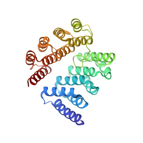
    Deoxyhypusine hydroxylase (DOHH) is a non-heme diiron enzyme involved in the posttranslational modification of a critical lysine residue of eukaryotic translation initiation factor 5A (eIF-5A) to yield the unusual amino acid residue hypusine. This modification is essential for the role of eIF-5A in translation and in nuclear export of a group of specific mRNAs. The diiron center of human DOHH (hDOHH) forms a peroxo-diiron(III) intermediate (hDOHHperoxo) when its reduced form reacts with O2. hDOHHperoxo has a lifetime exceeding that of the peroxo intermediates of other diiron enzymes by several orders of magnitude. Here we report the 1.7-Å crystal structures of hDOHHperoxo and a complex with glycerol. The structure of hDOHHperoxo reveals the presence of a μ-1,2-peroxo-diiron(III) species at the active site. Augmented by UV/Vis and Mössbauer spectroscopic studies, the crystal structures offer explanations for the extreme longevity of hDOHHperoxo and illustrate how the enzyme specifically recognizes its only substrate, deoxyhypusine-eIF-5A.


  • Organizational Affiliation

    Institute of Biochemistry, Center for Structural and Cell Biology in Medicine, University of Lübeck, Ratzeburger Allee 160, 23538 Lübeck, Germany.


Macromolecules
Find similar proteins by:  (by identity cutoff)  |  3D Structure
Entity ID: 1
MoleculeChains Sequence LengthOrganismDetailsImage
DEOXYHYPUSINE HYDROXYLASE
A, B
294Homo sapiensMutation(s): 0 
EC: 1.14.99.29
UniProt & NIH Common Fund Data Resources
Find proteins for Q9BU89 (Homo sapiens)
Explore Q9BU89 
Go to UniProtKB:  Q9BU89
PHAROS:  Q9BU89
GTEx:  ENSG00000129932 
Entity Groups  
Sequence Clusters30% Identity50% Identity70% Identity90% Identity95% Identity100% Identity
UniProt GroupQ9BU89
Sequence Annotations
Expand
  • Reference Sequence
Small Molecules
Ligands 3 Unique
IDChains Name / Formula / InChI Key2D Diagram3D Interactions
GAI
Query on GAI

Download Ideal Coordinates CCD File 
F [auth A],
G [auth A],
H [auth A],
I [auth A],
M [auth B]
GUANIDINE
C H5 N3
ZRALSGWEFCBTJO-UHFFFAOYSA-N
FE
Query on FE

Download Ideal Coordinates CCD File 
C [auth A],
D [auth A],
J [auth B],
K [auth B]
FE (III) ION
Fe
VTLYFUHAOXGGBS-UHFFFAOYSA-N
PER
Query on PER

Download Ideal Coordinates CCD File 
E [auth A],
L [auth B]
PEROXIDE ION
O2
ANAIPYUSIMHBEL-UHFFFAOYSA-N
Experimental Data & Validation

Experimental Data

  • Method: X-RAY DIFFRACTION
  • Resolution: 1.70 Å
  • R-Value Free: 0.246 
  • R-Value Work: 0.203 
  • R-Value Observed: 0.205 
  • Space Group: P 1 21 1
Unit Cell:
Length ( Å )Angle ( ˚ )
a = 45.933α = 90
b = 48.636β = 99.27
c = 124.087γ = 90
Software Package:
Software NamePurpose
REFMACrefinement
AutoPROCdata reduction
SCALAdata scaling
MOLREPphasing

Structure Validation

View Full Validation Report


Entry History 

Deposition Data

Revision History  (Full details and data files)

  • Version 1.0: 2015-04-15
    Type: Initial release
  • Version 1.1: 2015-04-22
    Changes: Database references
  • Version 1.2: 2015-05-27
    Changes: Database references