Photo-Oxidation of Tyrosine in a Bio-Engineered Bacterioferritin 'Reaction Centre'-A Protein Model for Artificial Photosynthesis.
Hingorani, K., Pace, R., Whitney, S., Murray, J.W., Smith, P., Cheah, M.H., Wydrzynski, T., Hillier, W.(2014) Biochim Biophys Acta 1837: 1821
- PubMed: 25107631
- DOI: https://doi.org/10.1016/j.bbabio.2014.07.019
- Primary Citation of Related Structures:
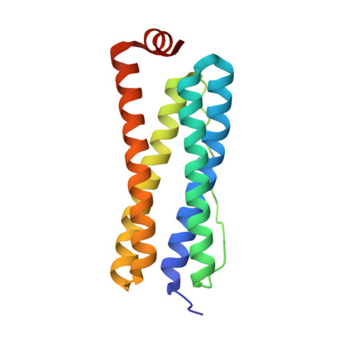
4CVP, 4CVR, 4CVS, 4CVT - PubMed Abstract:
The photosynthetic reaction centre (RC) is central to the conversion of solar energy into chemical energy and is a model for bio-mimetic engineering approaches to this end. We describe bio-engineering of a Photosystem II (PSII) RC inspired peptide model, building on our earlier studies. A non-photosynthetic haem containing bacterioferritin (BFR) from Escherichia coli that expresses as a homodimer was used as a protein scaffold, incorporating redox-active cofactors mimicking those of PSII. Desirable properties include: a di-nuclear metal binding site which provides ligands for bivalent metals, a hydrophobic pocket at the dimer interface which can bind a photosensitive porphyrin and presence of tyrosine residues proximal to the bound cofactors, which can be utilised as efficient electron-tunnelling intermediates. Light-induced electron transfer from proximal tyrosine residues to the photo-oxidised ZnCe6(•+), in the modified BFR reconstituted with both ZnCe6 and Mn(II), is presented. Three site-specific tyrosine variants (Y25F, Y58F and Y45F) were made to localise the redox-active tyrosine in the engineered system. The results indicate that: presence of bound Mn(II) is necessary to observe tyrosine oxidation in all BFR variants; Y45 the most important tyrosine as an immediate electron donor to the oxidised ZnCe6(•+) and that Y25 and Y58 are both redox-active in this system, but appear to function interchangebaly. High-resolution (2.1Å) crystal structures of the tyrosine variants show that there are no mutation-induced effects on the overall 3-D structure of the protein. Small effects are observed in the Y45F variant. Here, the BFR-RC represents a protein model for artificial photosynthesis.
Organizational Affiliation:
Building 134, Linnaeus Way, Research School of Biology, The Australian National University, ACT 0200, Australia. Electronic address: kastoori.hingorani@anu.edu.au.