4COK

Functional and Structural Characterization of Pyruvate decarboxylase from Gluconoacetobacter diazotrophicus


Experimental Data Snapshot

  • Method: X-RAY DIFFRACTION
  • Resolution: 1.69 Å
  • R-Value Free: 0.163 
  • R-Value Work: 0.132 
  • R-Value Observed: 0.133 

wwPDB Validation   3D Report Full Report


Ligand Structure Quality Assessment 


This is version 1.2 of the entry. See complete history


Literature

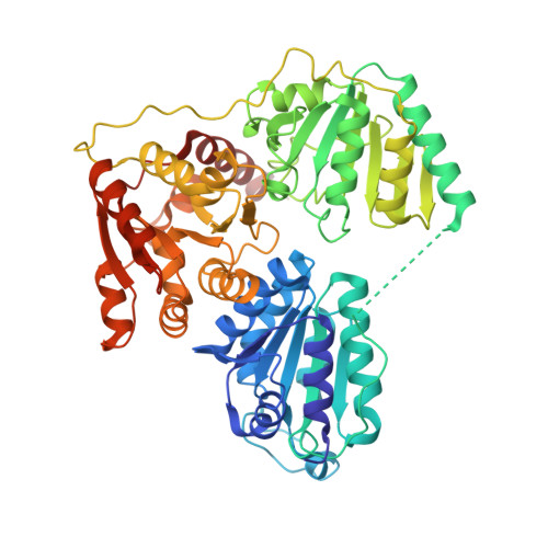
Structure and Functional Characterization of Pyruvate Decarboxylase from Gluconacetobacter Diazotrophicus.

Van Zyl, L.J.Schubert, W.Tuffin, M.I.Cowan, D.A.

(2014) BMC Struct Biol 14: 21

  • DOI: https://doi.org/10.1186/s12900-014-0021-1
  • Primary Citation of Related Structures:  
    4COK

  • PubMed Abstract: 

    Bacterial pyruvate decarboxylases (PDC) are rare. Their role in ethanol production and in bacterially mediated ethanologenic processes has, however, ensured a continued and growing interest. PDCs from Zymomonas mobilis (ZmPDC), Zymobacter palmae (ZpPDC) and Sarcina ventriculi (SvPDC) have been characterized and ZmPDC has been produced successfully in a range of heterologous hosts. PDCs from the Acetobacteraceae and their role in metabolism have not been characterized to the same extent. Examples include Gluconobacter oxydans (GoPDC), G. diazotrophicus (GdPDC) and Acetobacter pasteutrianus (ApPDC). All of these organisms are of commercial importance.


  • Organizational Affiliation

    Institute for Microbial Biotechnology and Metagenomics (IMBM), University of the Western Cape, Robert Sobukwe Road, Bellville, Cape Town, South Africa. vanzyllj@gmail.com.


Macromolecules
Find similar proteins by:  (by identity cutoff)  |  3D Structure
Entity ID: 1
MoleculeChains Sequence LengthOrganismDetailsImage
PYRUVATE DECARBOXYLASE
A, B
558Gluconacetobacter diazotrophicus PA1 5Mutation(s): 0 
EC: 4.1.1.1
UniProt
Find proteins for A9H275 (Gluconacetobacter diazotrophicus (strain ATCC 49037 / DSM 5601 / CCUG 37298 / CIP 103539 / LMG 7603 / PAl5))
Explore A9H275 
Go to UniProtKB:  A9H275
Entity Groups  
Sequence Clusters30% Identity50% Identity70% Identity90% Identity95% Identity100% Identity
UniProt GroupA9H275
Sequence Annotations
Expand
  • Reference Sequence
Experimental Data & Validation

Experimental Data

  • Method: X-RAY DIFFRACTION
  • Resolution: 1.69 Å
  • R-Value Free: 0.163 
  • R-Value Work: 0.132 
  • R-Value Observed: 0.133 
  • Space Group: C 1 2 1
Unit Cell:
Length ( Å )Angle ( ˚ )
a = 129.07α = 90
b = 140.96β = 125.78
c = 91.06γ = 90
Software Package:
Software NamePurpose
REFMACrefinement
MOSFLMdata reduction
SCALAdata scaling
MOLREPphasing

Structure Validation

View Full Validation Report


Ligand Structure Quality Assessment 


Entry History 

Deposition Data

Revision History  (Full details and data files)

  • Version 1.0: 2014-10-01
    Type: Initial release
  • Version 1.1: 2015-04-08
    Changes: Database references
  • Version 1.2: 2023-12-20
    Changes: Data collection, Database references, Derived calculations, Other, Refinement description