4C2U

Crystal structure of Deinococcus radiodurans UvrD in complex with DNA, Form 1


Experimental Data Snapshot

  • Method: X-RAY DIFFRACTION
  • Resolution: 2.55 Å
  • R-Value Free: 0.267 
  • R-Value Work: 0.211 
  • R-Value Observed: 0.214 

wwPDB Validation   3D Report Full Report


Ligand Structure Quality Assessment 


This is version 1.1 of the entry. See complete history


Literature

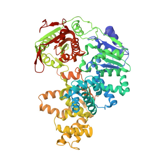
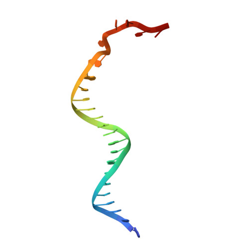
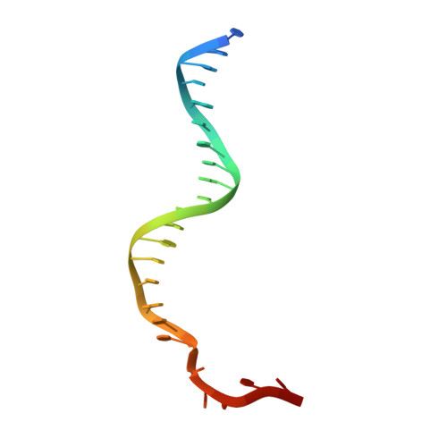
Structural and Mechanistic Insight Into DNA Unwinding by Deinococcus Radiodurans Uvrd.

Stelter, M.Acajjaoui, S.Mcsweeney, S.Timmins, J.

(2013) PLoS One 8: 77364

  • DOI: https://doi.org/10.1371/journal.pone.0077364
  • Primary Citation of Related Structures:  
    4C2T, 4C2U, 4C30

  • PubMed Abstract: 

    DNA helicases are responsible for unwinding the duplex DNA, a key step in many biological processes. UvrD is a DNA helicase involved in several DNA repair pathways. We report here crystal structures of Deinococcus radiodurans UvrD (drUvrD) in complex with DNA in different nucleotide-free and bound states. These structures provide us with three distinct snapshots of drUvrD in action and for the first time trap a DNA helicase undergoing a large-scale spiral movement around duplexed DNA. Our structural data also improve our understanding of the molecular mechanisms that regulate DNA unwinding by Superfamily 1A (SF1A) helicases. Our biochemical data reveal that drUvrD is a DNA-stimulated ATPase, can translocate along ssDNA in the 3'-5' direction and shows ATP-dependent 3'-5', and surprisingly also, 5'-3' helicase activity. Interestingly, we find that these translocase and helicase activities of drUvrD are modulated by the ssDNA binding protein. Analysis of drUvrD mutants indicate that the conserved β-hairpin structure of drUvrD that functions as a separation pin is critical for both drUvrD's 3'-5' and 5'-3' helicase activities, whereas the GIG motif of drUvrD involved in binding to the DNA duplex is essential for the 5'-3' helicase activity only. These special features of drUvrD may reflect its involvement in a wide range of DNA repair processes in vivo.


  • Organizational Affiliation

    Structural Biology Group, European Synchrotron Radiation Facility, Grenoble, France ; University Grenoble Alpes, Institut de Biologie structurale, Grenoble, France ; Centre National de la Recherche Scientifique, Institut de Biologie structurale, Grenoble, France ; Commissariat à l'énergie atomique et aux énergies alternatives, Département du Science du Vivant, Institut de Biologie structurale, Grenoble, France.


Macromolecules

Find similar proteins by:  (by identity cutoff)  |  3D Structure
Entity ID: 1
MoleculeChains Sequence LengthOrganismDetailsImage
DNA HELICASE IIA,
B [auth D]
665Deinococcus radioduransMutation(s): 0 
EC: 3.6.4.12
UniProt
Find proteins for Q9RTI9 (Deinococcus radiodurans (strain ATCC 13939 / DSM 20539 / JCM 16871 / CCUG 27074 / LMG 4051 / NBRC 15346 / NCIMB 9279 / VKM B-1422 / R1))
Explore Q9RTI9 
Go to UniProtKB:  Q9RTI9
Entity Groups  
Sequence Clusters30% Identity50% Identity70% Identity90% Identity95% Identity100% Identity
UniProt GroupQ9RTI9
Sequence Annotations
Expand
  • Reference Sequence

Find similar nucleic acids by:  Sequence   |   3D Structure  

Entity ID: 2
MoleculeChains LengthOrganismImage
REV25C [auth X]25synthetic construct
Sequence Annotations
Expand
  • Reference Sequence

Find similar nucleic acids by:  Sequence   |   3D Structure  

Entity ID: 3
MoleculeChains LengthOrganismImage
FOR25D [auth Y]25synthetic construct
Sequence Annotations
Expand
  • Reference Sequence
Small Molecules
Ligands 5 Unique
IDChains Name / Formula / InChI Key2D Diagram3D Interactions
ANP
Query on ANP

Download Ideal Coordinates CCD File 
E [auth A],
K [auth D]
PHOSPHOAMINOPHOSPHONIC ACID-ADENYLATE ESTER
C10 H17 N6 O12 P3
PVKSNHVPLWYQGJ-KQYNXXCUSA-N
GOL
Query on GOL

Download Ideal Coordinates CCD File 
H [auth A],
N [auth D]
GLYCEROL
C3 H8 O3
PEDCQBHIVMGVHV-UHFFFAOYSA-N
NO3
Query on NO3

Download Ideal Coordinates CCD File 
G [auth A],
I [auth A],
M [auth D],
O [auth D]
NITRATE ION
N O3
NHNBFGGVMKEFGY-UHFFFAOYSA-N
MG
Query on MG

Download Ideal Coordinates CCD File 
F [auth A],
L [auth D]
MAGNESIUM ION
Mg
JLVVSXFLKOJNIY-UHFFFAOYSA-N
NA
Query on NA

Download Ideal Coordinates CCD File 
J [auth A],
P [auth D]
SODIUM ION
Na
FKNQFGJONOIPTF-UHFFFAOYSA-N
Experimental Data & Validation

Experimental Data

  • Method: X-RAY DIFFRACTION
  • Resolution: 2.55 Å
  • R-Value Free: 0.267 
  • R-Value Work: 0.211 
  • R-Value Observed: 0.214 
  • Space Group: P 21 21 21
Unit Cell:
Length ( Å )Angle ( ˚ )
a = 67.572α = 90
b = 67.448β = 90
c = 386.04γ = 90
Software Package:
Software NamePurpose
REFMACrefinement
SCALAdata scaling
MrBUMPphasing

Structure Validation

View Full Validation Report


Ligand Structure Quality Assessment 


Entry History 

Deposition Data

Revision History  (Full details and data files)

  • Version 1.0: 2013-10-30
    Type: Initial release
  • Version 1.1: 2023-12-20
    Changes: Data collection, Database references, Derived calculations, Other, Refinement description