4C08

Crystal structure of M. musculus protein arginine methyltransferase PRMT6 with CaCl2 at 1.34 Angstroms


Experimental Data Snapshot

  • Method: X-RAY DIFFRACTION
  • Resolution: 1.34 Å
  • R-Value Free: 0.164 
  • R-Value Work: 0.141 
  • R-Value Observed: 0.142 

wwPDB Validation   3D Report Full Report


This is version 2.0 of the entry. See complete history


Literature

Functional Insights from High Resolution Structures of Mouse Protein Arginine Methyltransferase 6.

Bonnefond, L.Stojko, J.Mailliot, J.Troffer-Charlier, N.Cura, V.Wurtz, J.M.Cianferani, S.Cavarelli, J.

(2015) J Struct Biol 191: 175

  • DOI: https://doi.org/10.1016/j.jsb.2015.06.017
  • Primary Citation of Related Structures:  
    4C03, 4C04, 4C05, 4C06, 4C07, 4C08

  • PubMed Abstract: 

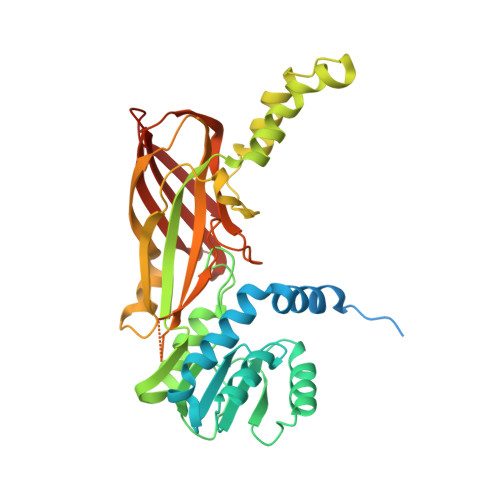
    PRMT6 is a protein arginine methyltransferase involved in transcriptional regulation, human immunodeficiency virus pathogenesis, DNA base excision repair, and cell cycle progression. Like other PRMTs, PRMT6 is overexpressed in several cancer types and is therefore considered as a potential anti-cancer drug target. In the present study, we described six crystal structures of PRMT6 from Mus musculus, solved and refined at 1.34 Å for the highest resolution structure. The crystal structures revealed that the folding of the helix αX is required to stabilize a productive active site before methylation of the bound peptide can occur. In the absence of cofactor, metal cations can be found in the catalytic pocket at the expected position of the guanidinium moiety of the target arginine substrate. Using mass spectrometry under native conditions, we show that PRMT6 dimer binds two cofactor and a single H4 peptide molecules. Finally, we characterized a new site of in vitro automethylation of mouse PRMT6 at position 7.


  • Organizational Affiliation

    Département de Biologie Structurale Intégrative, Institut de Génétique et de Biologie Moléculaire et Cellulaire (IGBMC), Université de Strasbourg, CNRS UMR7104, INSERM U964, 1 rue Laurent Fries, Illkirch, F-67404, France.


Macromolecules
Find similar proteins by:  (by identity cutoff)  |  3D Structure
Entity ID: 1
MoleculeChains Sequence LengthOrganismDetailsImage
PROTEIN ARGININE N-METHYLTRANSFERASE 6382Mus musculusMutation(s): 1 
EC: 2.1.1 (PDB Primary Data), 2.1.1.125 (PDB Primary Data)
UniProt & NIH Common Fund Data Resources
Find proteins for Q6NZB1 (Mus musculus)
Explore Q6NZB1 
Go to UniProtKB:  Q6NZB1
IMPC:  MGI:2139971
Entity Groups  
Sequence Clusters30% Identity50% Identity70% Identity90% Identity95% Identity100% Identity
UniProt GroupQ6NZB1
Sequence Annotations
Expand
  • Reference Sequence
Experimental Data & Validation

Experimental Data

  • Method: X-RAY DIFFRACTION
  • Resolution: 1.34 Å
  • R-Value Free: 0.164 
  • R-Value Work: 0.141 
  • R-Value Observed: 0.142 
  • Space Group: I 41
Unit Cell:
Length ( Å )Angle ( ˚ )
a = 79.325α = 90
b = 79.325β = 90
c = 118.411γ = 90
Software Package:
Software NamePurpose
PHENIXrefinement
XDSdata reduction
Aimlessdata scaling
PHASERphasing

Structure Validation

View Full Validation Report


Entry History 

Deposition Data

Revision History  (Full details and data files)

  • Version 1.0: 2014-07-30
    Type: Initial release
  • Version 1.1: 2015-07-01
    Changes: Database references
  • Version 1.2: 2015-08-12
    Changes: Database references
  • Version 2.0: 2023-12-20
    Changes: Atomic model, Data collection, Database references, Derived calculations, Other, Refinement description