4BXE

CRYSTAL STRUCTURE OF AMPDH3 FROM PSEUDOMONAS AERUGINOSA IN COMPLEX WITH ANHYDROMURAMIC PENTAPEPTIDE


Experimental Data Snapshot

  • Method: X-RAY DIFFRACTION
  • Resolution: 2.95 Å
  • R-Value Free: 0.266 
  • R-Value Work: 0.203 
  • R-Value Observed: 0.206 

wwPDB Validation   3D Report Full Report


This is version 2.0 of the entry. See complete history


Literature

Cell-Wall Remodeling by the Zinc-Protease Ampdh3 from Pseudomonas Aeruginosa.

Lee, M.Artola-Recolons, C.Carrasco-Lopez, C.Martinez-Caballero, S.Hesek, D.Spink, E.Lastochkin, E.Zhang, W.Hellman, L.M.Boggess, B.Hermoso, J.A.Mobashery, S.

(2013) J Am Chem Soc 135: 12604

  • DOI: https://doi.org/10.1021/ja407445x
  • Primary Citation of Related Structures:  
    4BXE, 4BXJ

  • PubMed Abstract: 

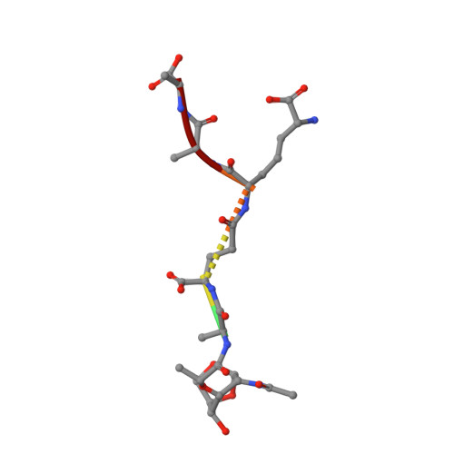
    Bacterial cell wall is a polymer of considerable complexity that is in constant equilibrium between synthesis and recycling. AmpDh3 is a periplasmic zinc protease of Pseudomonas aeruginosa , which is intimately involved in cell-wall remodeling. We document the hydrolytic reactions that this enzyme performs on the cell wall. The process removes the peptide stems from the peptidoglycan, the major constituent of the cell wall. We document that the majority of the reactions of this enzyme takes place on the polymeric insoluble portion of the cell wall, as opposed to the fraction that is released from it. We show that AmpDh3 is tetrameric both in crystals and in solution. Based on the X-ray structures of the enzyme in complex with two synthetic cell-wall-based ligands, we present for the first time a model for a multivalent anchoring of AmpDh3 onto the cell wall, which lends itself to its processive remodeling.


  • Organizational Affiliation

    Department of Chemistry and Biochemistry, University of Notre Dame, Notre Dame, Indiana 46556, USA.


Macromolecules
Find similar proteins by:  (by identity cutoff)  |  3D Structure
Entity ID: 1
MoleculeChains Sequence LengthOrganismDetailsImage
AMPDH3
A, B
255Pseudomonas aeruginosa PAO1Mutation(s): 0 
UniProt
Find proteins for Q9I5D1 (Pseudomonas aeruginosa (strain ATCC 15692 / DSM 22644 / CIP 104116 / JCM 14847 / LMG 12228 / 1C / PRS 101 / PAO1))
Explore Q9I5D1 
Go to UniProtKB:  Q9I5D1
Entity Groups  
Sequence Clusters30% Identity50% Identity70% Identity90% Identity95% Identity100% Identity
UniProt GroupQ9I5D1
Sequence Annotations
Expand
  • Reference Sequence

Find similar proteins by:  Sequence   |   3D Structure  

Entity ID: 2
MoleculeChains Sequence LengthOrganismDetailsImage
ANHYDROMURAMIC PEPTIDE
C, D
6synthetic constructMutation(s): 0 
Sequence Annotations
Expand
  • Reference Sequence
Experimental Data & Validation

Experimental Data

  • Method: X-RAY DIFFRACTION
  • Resolution: 2.95 Å
  • R-Value Free: 0.266 
  • R-Value Work: 0.203 
  • R-Value Observed: 0.206 
  • Space Group: P 41 21 2
Unit Cell:
Length ( Å )Angle ( ˚ )
a = 100.94α = 90
b = 100.94β = 90
c = 165.25γ = 90
Software Package:
Software NamePurpose
PHENIXmodel building
SCALAdata scaling
iMOSFLMphasing
SCALAphasing
MOLREPphasing
PHENIXphasing
PHENIXrefinement

Structure Validation

View Full Validation Report


Entry History 

Deposition Data

Revision History  (Full details and data files)

  • Version 1.0: 2013-10-09
    Type: Initial release
  • Version 1.1: 2013-10-16
    Changes: Atomic model, Structure summary
  • Version 2.0: 2023-11-15
    Changes: Atomic model, Data collection, Database references, Derived calculations, Other