4BPG

Crystal structure of Bacillus subtilis DltC


Experimental Data Snapshot

  • Method: X-RAY DIFFRACTION
  • Resolution: 2.20 Å
  • R-Value Free: 0.237 
  • R-Value Work: 0.209 
  • R-Value Observed: 0.210 

wwPDB Validation   3D Report Full Report


This is version 1.4 of the entry. See complete history


Literature

High-Resolution Structures of the D-Alanyl Carrier Protein (Dcp) Dltc from Bacillus Subtilis Reveal Equivalent Conformations of Apo- and Holo-Forms

Zimmermann, S.Pfennig, S.Neumann, P.Yonus, H.Weininger, U.Kovermann, M.Balbach, J.Stubbs, M.T.

(2015) FEBS Lett 589: 2283

  • DOI: https://doi.org/10.1016/j.febslet.2015.07.008
  • Primary Citation of Related Structures:  
    4BPF, 4BPG, 4BPH

  • PubMed Abstract: 

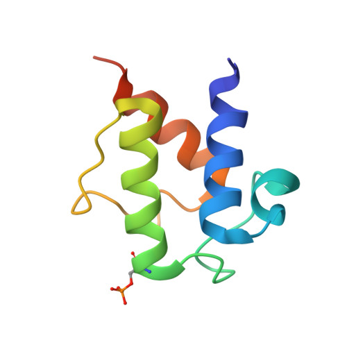
    D-Alanylation of lipoteichoic acids plays an important role in modulating the properties of Gram-positive bacteria cell walls. The D-alanyl carrier protein DltC from Bacillus subtilis has been solved in apo- and two cofactor-modified holo-forms, whereby the entire phosphopantetheine moiety is defined in one. The atomic resolution of the apo-structure allows delineation of alternative conformations within the hydrophobic core of the 78 residue four helix bundle. In contrast to previous reports for a peptidyl carrier protein from a non-ribosomal peptide synthetase, no obvious structural differences between apo- and holo-DltC forms are observed. Solution NMR spectroscopy confirms these findings and demonstrates in addition that the two forms exhibit similar backbone dynamics on the ps-ns and ms timescales.


  • Organizational Affiliation

    Institut für Biochemie und Biotechnologie, Martin-Luther Universität Halle-Wittenberg, Kurt-Mothes Strasse 3, D-06120 Halle/Saale, Germany.


Macromolecules
Find similar proteins by:  (by identity cutoff)  |  3D Structure
Entity ID: 1
MoleculeChains Sequence LengthOrganismDetailsImage
D-ALANINE--POLY(PHOSPHORIBITOL) LIGASE SUBUNIT 2
A, B
86Bacillus subtilisMutation(s): 0 
EC: 6.1.1.13
UniProt
Find proteins for P39579 (Bacillus subtilis (strain 168))
Explore P39579 
Go to UniProtKB:  P39579
Entity Groups  
Sequence Clusters30% Identity50% Identity70% Identity90% Identity95% Identity100% Identity
UniProt GroupP39579
Sequence Annotations
Expand
  • Reference Sequence
Small Molecules
Modified Residues  1 Unique
IDChains TypeFormula2D DiagramParent
SEP
Query on SEP
A, B
L-PEPTIDE LINKINGC3 H8 N O6 PSER
Experimental Data & Validation

Experimental Data

  • Method: X-RAY DIFFRACTION
  • Resolution: 2.20 Å
  • R-Value Free: 0.237 
  • R-Value Work: 0.209 
  • R-Value Observed: 0.210 
  • Space Group: P 61 2 2
Unit Cell:
Length ( Å )Angle ( ˚ )
a = 72.68α = 90
b = 72.68β = 90
c = 110.308γ = 120
Software Package:
Software NamePurpose
PHENIXrefinement
SCALAdata scaling
PHASERphasing

Structure Validation

View Full Validation Report


Entry History 

Deposition Data

Revision History  (Full details and data files)

  • Version 1.0: 2014-06-11
    Type: Initial release
  • Version 1.1: 2015-07-22
    Changes: Database references, Other
  • Version 1.2: 2015-07-29
    Changes: Database references
  • Version 1.3: 2015-08-26
    Changes: Database references
  • Version 1.4: 2023-12-20
    Changes: Data collection, Database references, Derived calculations, Other, Refinement description