4BN4

Structure of human SIRT3 in complex with ADP-ribose


Experimental Data Snapshot

  • Method: X-RAY DIFFRACTION
  • Resolution: 1.30 Å
  • R-Value Free: 0.179 
  • R-Value Work: 0.138 
  • R-Value Observed: 0.140 

wwPDB Validation   3D Report Full Report


Ligand Structure Quality Assessment 


This is version 1.6 of the entry. See complete history


Literature

Structures of Human Sirtuin 3 Complexes with Adp-Ribose and with Carba-Nad+ and Srt1720: Binding Details and Inhibition Mechanism

Nguyen, G.T.T.Schaefer, S.Gertz, M.Weyand, M.Steegborn, C.

(2013) Acta Crystallogr D Biol Crystallogr 69: 1423

  • DOI: https://doi.org/10.1107/S0907444913015448
  • Primary Citation of Related Structures:  
    4BN4, 4BN5

  • PubMed Abstract: 

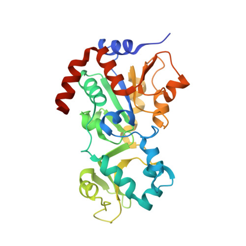
    Sirtuins are NAD(+)-dependent protein deacetylases that regulate metabolism and aging processes and are considered to be attractive therapeutic targets. Most available sirtuin modulators are little understood mechanistically, hindering their improvement. SRT1720 was initially described as an activator of human Sirt1, but it also potently inhibits human Sirt3. Here, the molecular mechanism of the inhibition of Sirt3 by SRT1720 is described. A crystal structure of Sirt3 in complex with SRT1720 and an NAD(+) analogue reveals that the compound partially occupies the acetyl-Lys binding site, thus explaining the reported competition with the peptide substrate. The compound packs against a hydrophobic protein patch and binds with its opposite surface to the NAD(+)  nicotinamide, resulting in an exceptionally tight sandwich-like interaction. The observed arrangement rationalizes the uncompetitive inhibition with NAD(+), and binding measurements confirm that the nicotinamide moiety of NAD(+) supports inhibitor binding. Consistently, no inhibitor is bound in a second crystal structure of Sirt3 that was solved complexed with ADP-ribose and crystallized in the presence of SRT1720. These results reveal a novel sirtuin inhibitor binding site and mechanism, and provide a structural basis for compound improvement.


  • Organizational Affiliation

    Department of Biochemistry, University of Bayreuth, Universitätsstrasse 30, 95445 Bayreuth, Germany.


Macromolecules
Find similar proteins by:  (by identity cutoff)  |  3D Structure
Entity ID: 1
MoleculeChains Sequence LengthOrganismDetailsImage
NAD-DEPENDENT PROTEIN DEACETYLASE SIRTUIN-3, MITOCHONDRIAL284Homo sapiensMutation(s): 0 
EC: 3.5.1
UniProt & NIH Common Fund Data Resources
Find proteins for Q9NTG7 (Homo sapiens)
Explore Q9NTG7 
Go to UniProtKB:  Q9NTG7
PHAROS:  Q9NTG7
GTEx:  ENSG00000142082 
Entity Groups  
Sequence Clusters30% Identity50% Identity70% Identity90% Identity95% Identity100% Identity
UniProt GroupQ9NTG7
Sequence Annotations
Expand
  • Reference Sequence
Small Molecules
Ligands 4 Unique
IDChains Name / Formula / InChI Key2D Diagram3D Interactions
AR6
Query on AR6

Download Ideal Coordinates CCD File 
D [auth A][(2R,3S,4R,5R)-5-(6-AMINOPURIN-9-YL)-3,4-DIHYDROXY-OXOLAN-2-YL]METHYL [HYDROXY-[[(2R,3S,4R,5S)-3,4,5-TRIHYDROXYOXOLAN-2-YL]METHOXY]PHOSPHORYL] HYDROGEN PHOSPHATE
C15 H23 N5 O14 P2
SRNWOUGRCWSEMX-ZQSHOCFMSA-N
OP2
Query on OP2

Download Ideal Coordinates CCD File 
E [auth A]2-[2-[2-[2-[2-(2-hydroxyethyloxy)ethoxy]ethoxy]ethoxy]ethoxy]ethanoic acid
C12 H24 O8
SXGGZTBEWZFLBZ-UHFFFAOYSA-N
ZN
Query on ZN

Download Ideal Coordinates CCD File 
B [auth A]ZINC ION
Zn
PTFCDOFLOPIGGS-UHFFFAOYSA-N
NA
Query on NA

Download Ideal Coordinates CCD File 
C [auth A]SODIUM ION
Na
FKNQFGJONOIPTF-UHFFFAOYSA-N
Experimental Data & Validation

Experimental Data

  • Method: X-RAY DIFFRACTION
  • Resolution: 1.30 Å
  • R-Value Free: 0.179 
  • R-Value Work: 0.138 
  • R-Value Observed: 0.140 
  • Space Group: P 21 21 21
Unit Cell:
Length ( Å )Angle ( ˚ )
a = 63.9α = 90
b = 66.67β = 90
c = 66.85γ = 90
Software Package:
Software NamePurpose
REFMACrefinement
XDSdata reduction
XSCALEdata scaling
PHASERphasing

Structure Validation

View Full Validation Report


Ligand Structure Quality Assessment 


Entry History 

Deposition Data

Revision History  (Full details and data files)

  • Version 1.0: 2013-06-26
    Type: Initial release
  • Version 1.1: 2013-07-24
    Changes: Atomic model, Other
  • Version 1.2: 2013-07-31
    Changes: Database references
  • Version 1.3: 2013-08-07
    Changes: Atomic model, Other
  • Version 1.4: 2013-08-14
    Changes: Atomic model, Other
  • Version 1.5: 2013-08-28
    Changes: Atomic model, Other
  • Version 1.6: 2023-12-20
    Changes: Data collection, Database references, Derived calculations, Other, Refinement description, Structure summary