Crystal Structure of 3-Hydroxybenzoate 6-Hydroxylase Uncovers Lipid-Assisted Flavoprotein Strategy for Regioselective Aromatic Hydroxylation
Montersino, S., Orru, R., Barendregt, A., Westphal, A.H., Van Duijn, E., Mattevi, A., Van Berkel, W.J.H.(2013) J Biol Chem 288: 26235
- PubMed: 23864660
- DOI: https://doi.org/10.1074/jbc.M113.479303
- PubMed Abstract:
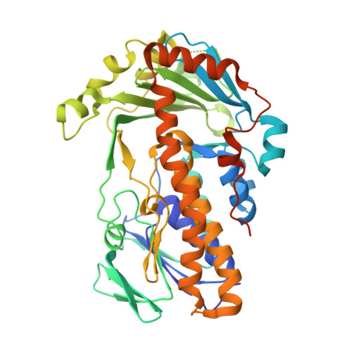
3-Hydroxybenzoate 6-hydroxylase (3HB6H) from Rhodococcus jostii RHA1 is a dimeric flavoprotein that catalyzes the NADH- and oxygen-dependent para-hydroxylation of 3-hydroxybenzoate to 2,5-dihydroxybenzoate. In this study, we report the crystal structure of 3HB6H as expressed in Escherichia coli. The overall fold of 3HB6H is similar to that of p-hydroxybenzoate hydroxylase and other flavoprotein aromatic hydroxylases. Unexpectedly, a lipid ligand is bound to each 3HB6H monomer. Mass spectral analysis identified the ligand as a mixture of phosphatidylglycerol and phosphatidylethanolamine. The fatty acid chains occupy hydrophobic channels that deeply penetrate into the interior of the substrate-binding domain of each subunit, whereas the hydrophilic part is exposed on the protein surface, connecting the dimerization domains via a few interactions. Most remarkably, the terminal part of a phospholipid acyl chain is directly involved in the substrate-binding site. Co-crystallized chloride ion and the crystal structure of the H213S variant with bound 3-hydroxybenzoate provide hints about oxygen activation and substrate hydroxylation. Essential roles are played by His-213 in catalysis and Tyr-105 in substrate binding. This phospholipid-assisted strategy to control regioselective aromatic hydroxylation is of relevance for optimization of flavin-dependent biocatalysts.
Organizational Affiliation:
From the Laboratory of Biochemistry, Wageningen University, 6703 HA Wageningen, The Netherlands.