4BJP

Crystal structure of E. coli penicillin binding protein 3


Experimental Data Snapshot

  • Method: X-RAY DIFFRACTION
  • Resolution: 2.50 Å
  • R-Value Free: 0.245 
  • R-Value Work: 0.199 
  • R-Value Observed: 0.201 

wwPDB Validation   3D Report Full Report


Ligand Structure Quality Assessment 


This is version 1.2 of the entry. See complete history


Literature

Crystal Structure of Penicillin-Binding Protein 3 (Pbp3) from Escherichia Coli.

Sauvage, E.Derouaux, A.Fraipont, C.Joris, M.Herman, R.Rocaboy, M.Schloesser, M.Dumas, J.Kerff, F.Nguyen-Disteche, M.Charlier, P.

(2014) PLoS One 9: 98042

  • DOI: https://doi.org/10.1371/journal.pone.0098042
  • Primary Citation of Related Structures:  
    4BJP, 4BJQ

  • PubMed Abstract: 

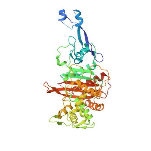
    In Escherichia coli, penicillin-binding protein 3 (PBP3), also known as FtsI, is a central component of the divisome, catalyzing cross-linking of the cell wall peptidoglycan during cell division. PBP3 is mainly periplasmic, with a 23 residues cytoplasmic tail and a single transmembrane helix. We have solved the crystal structure of a soluble form of PBP3 (PBP3(57-577)) at 2.5 Å revealing the two modules of high molecular weight class B PBPs, a carboxy terminal module exhibiting transpeptidase activity and an amino terminal module of unknown function. To gain additional insight, the PBP3 Val88-Ser165 subdomain (PBP3(88-165)), for which the electron density is poorly defined in the PBP3 crystal, was produced and its structure solved by SAD phasing at 2.1 Å. The structure shows a three dimensional domain swapping with a β-strand of one molecule inserted between two strands of the paired molecule, suggesting a possible role in PBP3(57-577) dimerization.


  • Organizational Affiliation

    Centre d'Ingénierie des Protéines, Université de Liège, Institut de Physique B5a et Institut de Chimie B6a, Sart Tilman, Liège, Belgium.


Macromolecules
Find similar proteins by:  (by identity cutoff)  |  3D Structure
Entity ID: 1
MoleculeChains Sequence LengthOrganismDetailsImage
PENICILLIN BINDING PROTEIN TRANSPEPTIDASE DOMAIN PROTEIN511Escherichia coliMutation(s): 0 
EC: 2.4.1.129
Entity Groups  
Sequence Clusters30% Identity50% Identity70% Identity90% Identity95% Identity100% Identity
Sequence Annotations
Expand
  • Reference Sequence
Small Molecules
Ligands 5 Unique
IDChains Name / Formula / InChI Key2D Diagram3D Interactions
CXS
Query on CXS

Download Ideal Coordinates CCD File 
F [auth A],
G [auth A],
H [auth A]
3-CYCLOHEXYL-1-PROPYLSULFONIC ACID
C9 H19 N O3 S
PJWWRFATQTVXHA-UHFFFAOYSA-N
SO4
Query on SO4

Download Ideal Coordinates CCD File 
B [auth A],
C [auth A],
D [auth A],
E [auth A]
SULFATE ION
O4 S
QAOWNCQODCNURD-UHFFFAOYSA-L
GOL
Query on GOL

Download Ideal Coordinates CCD File 
I [auth A]
J [auth A]
K [auth A]
L [auth A]
M [auth A]
I [auth A],
J [auth A],
K [auth A],
L [auth A],
M [auth A],
N [auth A],
O [auth A],
P [auth A],
Q [auth A],
R [auth A],
S [auth A]
GLYCEROL
C3 H8 O3
PEDCQBHIVMGVHV-UHFFFAOYSA-N
EDO
Query on EDO

Download Ideal Coordinates CCD File 
AA [auth A]
BA [auth A]
CA [auth A]
T [auth A]
U [auth A]
AA [auth A],
BA [auth A],
CA [auth A],
T [auth A],
U [auth A],
V [auth A],
W [auth A],
X [auth A],
Y [auth A],
Z [auth A]
1,2-ETHANEDIOL
C2 H6 O2
LYCAIKOWRPUZTN-UHFFFAOYSA-N
CL
Query on CL

Download Ideal Coordinates CCD File 
DA [auth A]CHLORIDE ION
Cl
VEXZGXHMUGYJMC-UHFFFAOYSA-M
Experimental Data & Validation

Experimental Data

  • Method: X-RAY DIFFRACTION
  • Resolution: 2.50 Å
  • R-Value Free: 0.245 
  • R-Value Work: 0.199 
  • R-Value Observed: 0.201 
  • Space Group: P 61 2 2
Unit Cell:
Length ( Å )Angle ( ˚ )
a = 119.025α = 90
b = 119.025β = 90
c = 139.251γ = 120
Software Package:
Software NamePurpose
REFMACrefinement
MOSFLMdata reduction
SCALAdata scaling
PHASERphasing

Structure Validation

View Full Validation Report


Ligand Structure Quality Assessment 


Entry History 

Deposition Data

Revision History  (Full details and data files)

  • Version 1.0: 2014-05-07
    Type: Initial release
  • Version 1.1: 2014-06-18
    Changes: Database references
  • Version 1.2: 2023-12-20
    Changes: Data collection, Database references, Derived calculations, Other, Refinement description