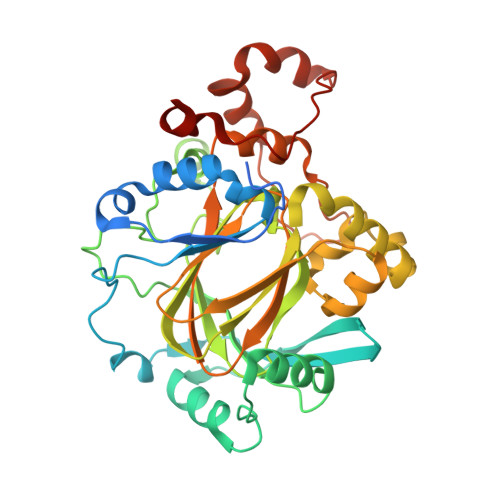
4BIS

JMJD2A COMPLEXED WITH 8-HYDROXYQUINOLINE-4-CARBOXYLIC ACID


Experimental Data Snapshot

  • Method: X-RAY DIFFRACTION
  • Resolution: 2.49 Å
  • R-Value Free: 0.224 
  • R-Value Work: 0.193 
  • R-Value Observed: 0.194 

wwPDB Validation   3D Report Full Report


Ligand Structure Quality Assessment 


This is version 1.4 of the entry. See complete history


Literature

5-Carboxy-8-hydroxyquinoline is a Broad Spectrum 2-Oxoglutarate Oxygenase Inhibitor which Causes Iron Translocation.

Hopkinson, R.J.Tumber, A.Yapp, C.Chowdhury, R.Aik, W.Che, K.H.Li, X.S.Kristensen, J.B.L.King, O.N.F.Chan, M.C.Yeoh, K.K.Choi, H.Walport, L.J.Thinnes, C.C.Bush, J.T.Lejeune, C.Rydzik, A.M.Rose, N.R.Bagg, E.A.McDonough, M.A.Krojer, T.Yue, W.W.Ng, S.S.Olsen, L.Brennan, P.E.Oppermann, U.Muller-Knapp, S.Klose, R.J.Ratcliffe, P.J.Schofield, C.J.Kawamura, A.

(2013) Chem Sci 4: 3110-3117

  • DOI: https://doi.org/10.1039/C3SC51122G
  • Primary Citation of Related Structures:  
    4BIO, 4BIS, 4JHT

  • PubMed Abstract: 

    2-Oxoglutarate and iron dependent oxygenases are therapeutic targets for human diseases. Using a representative 2OG oxygenase panel, we compare the inhibitory activities of 5-carboxy-8-hydroxyquinoline (IOX1) and 4-carboxy-8-hydroxyquinoline (4C8HQ) with that of two other commonly used 2OG oxygenase inhibitors, N -oxalylglycine (NOG) and 2,4-pyridinedicarboxylic acid (2,4-PDCA). The results reveal that IOX1 has a broad spectrum of activity, as demonstrated by the inhibition of transcription factor hydroxylases, representatives of all 2OG dependent histone demethylase subfamilies, nucleic acid demethylases and γ-butyrobetaine hydroxylase. Cellular assays show that, unlike NOG and 2,4-PDCA, IOX1 is active against both cytosolic and nuclear 2OG oxygenases without ester derivatisation. Unexpectedly, crystallographic studies on these oxygenases demonstrate that IOX1, but not 4C8HQ, can cause translocation of the active site metal, revealing a rare example of protein ligand-induced metal movement.


  • Organizational Affiliation

    Chemistry Research Laboratory, Department of Chemistry, University of Oxford, Mansfield Road, Oxford, OX1 3TA, U.K.


Macromolecules
Find similar proteins by:  (by identity cutoff)  |  3D Structure
Entity ID: 1
MoleculeChains Sequence LengthOrganismDetailsImage
LYSINE-SPECIFIC DEMETHYLASE 4A
A, B
381Homo sapiensMutation(s): 0 
EC: 1.14.11
UniProt & NIH Common Fund Data Resources
Find proteins for O75164 (Homo sapiens)
Explore O75164 
Go to UniProtKB:  O75164
PHAROS:  O75164
GTEx:  ENSG00000066135 
Entity Groups  
Sequence Clusters30% Identity50% Identity70% Identity90% Identity95% Identity100% Identity
UniProt GroupO75164
Sequence Annotations
Expand
  • Reference Sequence
Small Molecules
Ligands 5 Unique
IDChains Name / Formula / InChI Key2D Diagram3D Interactions
8HQ
Query on 8HQ

Download Ideal Coordinates CCD File 
F [auth A],
K [auth B]
8-hydroxyquinoline-4-carboxylic acid
C10 H7 N O3
KPTUKANGPPVLMD-UHFFFAOYSA-N
GOL
Query on GOL

Download Ideal Coordinates CCD File 
G [auth A]GLYCEROL
C3 H8 O3
PEDCQBHIVMGVHV-UHFFFAOYSA-N
ZN
Query on ZN

Download Ideal Coordinates CCD File 
D [auth A],
I [auth B]
ZINC ION
Zn
PTFCDOFLOPIGGS-UHFFFAOYSA-N
NI
Query on NI

Download Ideal Coordinates CCD File 
C [auth A],
H [auth B]
NICKEL (II) ION
Ni
VEQPNABPJHWNSG-UHFFFAOYSA-N
CL
Query on CL

Download Ideal Coordinates CCD File 
E [auth A],
J [auth B]
CHLORIDE ION
Cl
VEXZGXHMUGYJMC-UHFFFAOYSA-M
Binding Affinity Annotations 
IDSourceBinding Affinity
8HQ BindingDB:  4BIS IC50: 1700 (nM) from 1 assay(s)
Experimental Data & Validation

Experimental Data

  • Method: X-RAY DIFFRACTION
  • Resolution: 2.49 Å
  • R-Value Free: 0.224 
  • R-Value Work: 0.193 
  • R-Value Observed: 0.194 
  • Space Group: P 21 21 2
Unit Cell:
Length ( Å )Angle ( ˚ )
a = 100.69α = 90
b = 150.15β = 90
c = 57.45γ = 90
Software Package:
Software NamePurpose
PHENIXrefinement
XDSdata reduction
SCALAdata scaling
PHASERphasing

Structure Validation

View Full Validation Report


Ligand Structure Quality Assessment 


Entry History 

Deposition Data

Revision History  (Full details and data files)

  • Version 1.0: 2014-02-05
    Type: Initial release
  • Version 1.1: 2018-03-28
    Changes: Database references
  • Version 1.2: 2019-03-06
    Changes: Data collection, Experimental preparation
  • Version 1.3: 2019-05-08
    Changes: Data collection, Experimental preparation
  • Version 1.4: 2023-12-20
    Changes: Data collection, Database references, Derived calculations, Other, Refinement description