Superoxide Reductase from Giardia Intestinalis: Structural Characterization of the First Sor from a Eukaryotic Organism Shows an Iron Centre that is Highly Sensitive to Photoreduction
Bandeiras, T.M., Rodrigues, J.V., Sousa, C.M., Barradas, A.R., Pinho, F.G., Pinto, A.F., Teixeira, M., Matias, P.M., Romao, C.V.(2015) Acta Crystallogr D Biol Crystallogr 71: 2236
- PubMed: 26527141
- DOI: https://doi.org/10.1107/S1399004715015825
- Primary Citation of Related Structures:
4BGL, 4D7P - PubMed Abstract:
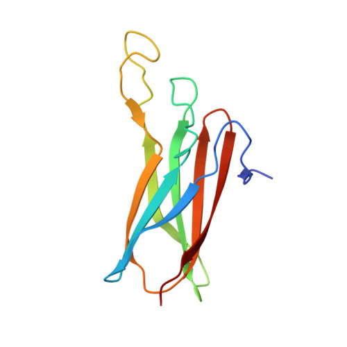
Superoxide reductase (SOR), which is commonly found in prokaryotic organisms, affords protection from oxidative stress by reducing the superoxide anion to hydrogen peroxide. The reaction is catalyzed at the iron centre, which is highly conserved among the prokaryotic SORs structurally characterized to date. Reported here is the first structure of an SOR from a eukaryotic organism, the protozoan parasite Giardia intestinalis (GiSOR), which was solved at 2.0 Å resolution. By collecting several diffraction data sets at 100 K from the same flash-cooled protein crystal using synchrotron X-ray radiation, photoreduction of the iron centre was observed. Reduction was monitored using an online UV-visible microspectrophotometer, following the decay of the 647 nm absorption band characteristic of the iron site in the glutamate-bound, oxidized state. Similarly to other 1Fe-SORs structurally characterized to date, the enzyme displays a tetrameric quaternary-structure arrangement. As a distinctive feature, the N-terminal loop of the protein, containing the characteristic EKHxP motif, revealed an unusually high flexibility regardless of the iron redox state. At variance with previous evidence collected by X-ray crystallography and Fourier transform infrared spectroscopy of prokaryotic SORs, iron reduction did not lead to dissociation of glutamate from the catalytic metal or other structural changes; however, the glutamate ligand underwent X-ray-induced chemical changes, revealing high sensitivity of the GiSOR active site to X-ray radiation damage.
Organizational Affiliation:
Instituto de Tecnologia Química e Biológica, António Xavier Universidade Nova de Lisboa, Avenida da República, 2780-157 Oeiras, Portugal.