4BER

Crystal structure of the Legionella pneumophila FIC domain-containing effector AnkX protein in complex with cytidine monophosphate


Experimental Data Snapshot

  • Method: X-RAY DIFFRACTION
  • Resolution: 2.60 Å
  • R-Value Free: 0.239 
  • R-Value Work: 0.206 
  • R-Value Observed: 0.207 

wwPDB Validation   3D Report Full Report


Ligand Structure Quality Assessment 


This is version 1.2 of the entry. See complete history


Literature

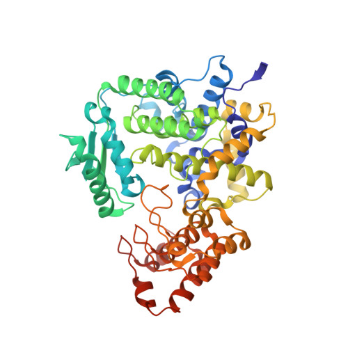
Structure of the Legionella Effector Ankx Reveals the Mechanism of Phosphocholine Transfer by the Fic Domain.

Campanacci, V.Mukherjee, S.Roy, C.R.Cherfils, J.

(2013) EMBO J 32: 1469

  • DOI: https://doi.org/10.1038/emboj.2013.82
  • Primary Citation of Related Structures:  
    4BEP, 4BER, 4BES, 4BET

  • PubMed Abstract: 

    The FIC motif and the eukaryotic-like ankyrin repeats are found in many bacterial type IV effectors, yet little is known about how these domains enable bacteria to modulate host cell functions. Bacterial FIC domains typically bind ATP and transfer adenosine monophosphate moiety onto target proteins. The ankyrin repeat-containing protein AnkX encoded by the intracellular pathogen Legionella pneumophila is unique in that its FIC domain binds to CDP-choline and transfers a phosphocholine residue onto proteins in the Rab1 GTPase family. By determining the structures of unbound AnkX and AnkX with bound CDP-choline, CMP/phosphocholine and CMP, we demonstrate that the orientation of substrate binding in relation to the catalytic FIC motif enables this protein to function as a phosphocholinating enzyme rather than a nucleotidyl transferase. Additionally, the structure reveals that the ankyrin repeats mediate scaffolding interactions that resemble those found in protein-protein interactions, but are unprecedented in intramolecular interactions. Together with phosphocholination experiments, our structures unify a general phosphoryl transferase mechanism common to all FIC enzymes that should be conserved from bacteria to human.


  • Organizational Affiliation

    Laboratoire d'Enzymologie et Biochimie Structurales, Centre de Recherche de Gif, CNRS, Gif-sur-Yvette, France.


Macromolecules
Find similar proteins by:  (by identity cutoff)  |  3D Structure
Entity ID: 1
MoleculeChains Sequence LengthOrganismDetailsImage
PHOSPHOCHOLINE TRANSFERASE ANKX
A, B
484Legionella pneumophilaMutation(s): 1 
EC: 2.7.1
UniProt
Find proteins for Q5ZXN6 (Legionella pneumophila subsp. pneumophila (strain Philadelphia 1 / ATCC 33152 / DSM 7513))
Explore Q5ZXN6 
Go to UniProtKB:  Q5ZXN6
Entity Groups  
Sequence Clusters30% Identity50% Identity70% Identity90% Identity95% Identity100% Identity
UniProt GroupQ5ZXN6
Sequence Annotations
Expand
  • Reference Sequence
Experimental Data & Validation

Experimental Data

  • Method: X-RAY DIFFRACTION
  • Resolution: 2.60 Å
  • R-Value Free: 0.239 
  • R-Value Work: 0.206 
  • R-Value Observed: 0.207 
  • Space Group: P 2 2 21
Unit Cell:
Length ( Å )Angle ( ˚ )
a = 58.33α = 90
b = 103.56β = 90
c = 224.1γ = 90
Software Package:
Software NamePurpose
BUSTERrefinement
XDSdata reduction
XDSdata scaling
PHASERphasing

Structure Validation

View Full Validation Report


Ligand Structure Quality Assessment 


Entry History 

Deposition Data

Revision History  (Full details and data files)

  • Version 1.0: 2013-04-24
    Type: Initial release
  • Version 1.1: 2013-05-29
    Changes: Database references
  • Version 1.2: 2023-12-20
    Changes: Data collection, Database references, Derived calculations, Other, Refinement description