4AUI

STRUCTURE AND FUNCTION OF THE PORB PORIN FROM DISSEMINATING N. GONORRHOEAE


Experimental Data Snapshot

  • Method: X-RAY DIFFRACTION
  • Resolution: 3.20 Å
  • R-Value Free: 0.268 
  • R-Value Work: 0.221 
  • R-Value Observed: 0.224 

wwPDB Validation   3D Report Full Report


Ligand Structure Quality Assessment 


This is version 1.4 of the entry. See complete history


Literature

Structure and Function of the Porb Porin from Disseminating Neisseria Gonorrhoeae.

Zeth, K.Kozjak-Pavlovic, V.Faulstich, M.Fraunholz, M.Hurwitz, R.Kepp, O.Rudel, T.

(2013) Biochem J 449: 631

  • DOI: https://doi.org/10.1042/BJ20121025
  • Primary Citation of Related Structures:  
    4AUI

  • PubMed Abstract: 

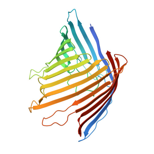
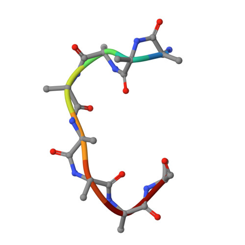
    The outer membrane of Gram-negative bacteria contains a large number of channel-forming proteins, porins, for the uptake of small nutrient molecules. Neisseria gonorrhoeae PorBIA (PorB of serotype A) are associated with disseminating diseases and mediate a rapid bacterial invasion into host cells in a phosphate-sensitive manner. To gain insights into this structure-function relationship we analysed PorBIA by X-ray crystallography in the presence of phosphate and ATP. The structure of PorBIA in the complex solved at a resolution of 3.3 Å (1 Å=0.1 nm) displays a surplus of positive charges inside the channel. ATP ligand-binding in the channel is co-ordinated by the positively charged residues of the channel interior. These residues ligate the aromatic, sugar and pyrophosphate moieties of the ligand. Two phosphate ions were observed in the structure, one of which clamped by two arginine residues (Arg92 and Arg124) localized at the extraplasmic channel exit. A short β-bulge in β2-strand together with the long L3 loop narrow the barrel diameter significantly and further support substrate specificity through hydrogen bond interactions. Interestingly the structure also comprised a small peptide as a remnant of a periplasmic protein which physically links porin molecules to the peptidoglycan network. To test the importance of Arg92 on bacterial invasion the residue was mutated. In vivo assays of bacteria carrying a R92S mutation confirmed the importance of this residue for host-cell invasion. Furthermore systematic sequence and structure comparisons of PorBIA from Neisseriaceae indicated Arg92 to be unique in disseminating N. gonorrhoeae thereby possibly distinguishing invasion-promoting porins from other neisserial porins.


  • Organizational Affiliation

    Max Planck Institute for Developmental Biology, Spemannstr. 35, 72076 Tübingen, Germany. kornelius.zeth@googlemail.com


Macromolecules
Find similar proteins by:  (by identity cutoff)  |  3D Structure
Entity ID: 1
MoleculeChains Sequence LengthOrganismDetailsImage
PORIN (PORB)
A, B, C
327Neisseria gonorrhoeaeMutation(s): 0 
Membrane Entity: Yes 
UniProt
Find proteins for Q51056 (Neisseria gonorrhoeae)
Explore Q51056 
Go to UniProtKB:  Q51056
Entity Groups  
Sequence Clusters30% Identity50% Identity70% Identity90% Identity95% Identity100% Identity
UniProt GroupQ51056
Sequence Annotations
Expand
  • Reference Sequence

Find similar proteins by:  Sequence   |   3D Structure  

Entity ID: 2
MoleculeChains Sequence LengthOrganismDetailsImage
POLY ALA
D, E, F
8Neisseria gonorrhoeaeMutation(s): 0 
Sequence Annotations
Expand
  • Reference Sequence
Small Molecules
Ligands 3 Unique
IDChains Name / Formula / InChI Key2D Diagram3D Interactions
ATP
Query on ATP

Download Ideal Coordinates CCD File 
G [auth A],
K [auth B],
N [auth C]
ADENOSINE-5'-TRIPHOSPHATE
C10 H16 N5 O13 P3
ZKHQWZAMYRWXGA-KQYNXXCUSA-N
PO4
Query on PO4

Download Ideal Coordinates CCD File 
H [auth A]
I [auth A]
L [auth B]
M [auth B]
O [auth C]
H [auth A],
I [auth A],
L [auth B],
M [auth B],
O [auth C],
P [auth C]
PHOSPHATE ION
O4 P
NBIIXXVUZAFLBC-UHFFFAOYSA-K
MG
Query on MG

Download Ideal Coordinates CCD File 
J [auth A]MAGNESIUM ION
Mg
JLVVSXFLKOJNIY-UHFFFAOYSA-N
Experimental Data & Validation

Experimental Data

  • Method: X-RAY DIFFRACTION
  • Resolution: 3.20 Å
  • R-Value Free: 0.268 
  • R-Value Work: 0.221 
  • R-Value Observed: 0.224 
  • Space Group: C 1 2 1
Unit Cell:
Length ( Å )Angle ( ˚ )
a = 114.05α = 90
b = 111.47β = 102.07
c = 88.34γ = 90
Software Package:
Software NamePurpose
PHENIXrefinement
XDSdata reduction
XSCALEdata scaling
MOLREPphasing

Structure Validation

View Full Validation Report


Ligand Structure Quality Assessment 


Entry History 

Deposition Data

Revision History  (Full details and data files)

  • Version 1.0: 2012-10-10
    Type: Initial release
  • Version 1.1: 2012-11-14
    Changes: Database references
  • Version 1.2: 2013-01-23
    Changes: Database references
  • Version 1.3: 2019-10-09
    Changes: Advisory, Data collection, Other, Source and taxonomy, Structure summary
  • Version 1.4: 2023-12-20
    Changes: Advisory, Data collection, Database references, Derived calculations, Refinement description