4AOC

crystal structure of BC2L-A Lectin from Burkolderia cenocepacia in complex with methyl-heptoside


Experimental Data Snapshot

  • Method: X-RAY DIFFRACTION
  • Resolution: 2.70 Å
  • R-Value Free: 0.253 
  • R-Value Work: 0.192 
  • R-Value Observed: 0.195 

wwPDB Validation   3D Report Full Report


Ligand Structure Quality Assessment 


This is version 1.4 of the entry. See complete history


Literature

Burkholderia Cenocepacia Lectin a Binding to Heptoses from the Bacterial Lipopolysaccharide.

Marchetti, R.Malinovska, L.Lameignere, E.Adamova, L.De Castro, C.Cioci, G.Stanetty, C.Kosma, P.Molinaro, A.Wimmerova, M.Imberty, A.Silipo, A.

(2012) Glycobiology 22: 1387

  • DOI: https://doi.org/10.1093/glycob/cws105
  • Primary Citation of Related Structures:  
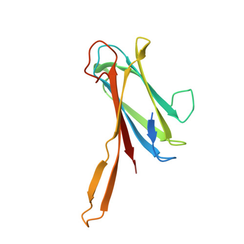
    4AOC

  • PubMed Abstract: 

    Bacteria from the Burkholderia cepacia complex (Bcc) cause highly contagious pneumonia among cystic fibrosis (CF) patients. Among them, Burkholderia cenocepacia is one of the most dangerous in the Bcc and is the most frequent cause of morbidity and mortality in CF patients. Indeed, it is responsible of "cepacia syndrome", a deadly exacerbation of infection, that is the main cause of poor outcomes in lung transplantation. Burkholderia cenocepacia produces several soluble lectins with specificity for fucosylated and mannosylated glycoconjugates. These lectins are present on the bacterial cell surface and it has been proposed that they bind to lipopolysaccharide epitopes. In this work, we report on the interaction of one B. cenocepacia lectin, BC2L-A, with heptose and other manno configured sugar residues. Saturation transfer difference NMR spectroscopy studies of BC2L-A with different mono- and disaccharides demonstrated the requirement of manno configuration with the hydroxyl or glycol group at C6 for the binding process. The crystal structure of BC2L-A complexed with the methyl-heptoside confirmed the location of the carbohydrate ring in the binding site and elucidated the orientation of the glycol tail, in agreement with NMR data. Titration calorimetry performed on monosaccharides, heptose disaccharides and bacterial heptose-containing oligosaccharides and polysaccharides confirmed that bacterial cell wall contains carbohydrate epitopes that can bind to BC2L-A. Additionally, the specific binding of fluorescent BC2L-A lectin on B. cenocepacia bacterial surface was demonstrated by microscopy.


  • Organizational Affiliation

    Dipartimento di Scienze Chimiche, Università di Napoli "Federico II", Italy.


Macromolecules
Find similar proteins by:  (by identity cutoff)  |  3D Structure
Entity ID: 1
MoleculeChains Sequence LengthOrganismDetailsImage
BC2L-A LECTIN
A, B, C, D, E
129Burkholderia orbicola AU 1054Mutation(s): 0 
UniProt
Find proteins for A0A0H2Y045 (Burkholderia orbicola (strain AU 1054))
Explore A0A0H2Y045 
Go to UniProtKB:  A0A0H2Y045
Entity Groups  
Sequence Clusters30% Identity50% Identity70% Identity90% Identity95% Identity100% Identity
UniProt GroupA0A0H2Y045
Sequence Annotations
Expand
  • Reference Sequence
Small Molecules
Ligands 3 Unique
IDChains Name / Formula / InChI Key2D Diagram3D Interactions
A1Q
Query on A1Q

Download Ideal Coordinates CCD File 
DA [auth E],
H [auth A],
M [auth B],
Q [auth C],
X [auth D]
methyl L-glycero-alpha-D-manno-heptopyranoside
C8 H16 O7
GJUAFBSAJCBGRU-IHKZFYOVSA-N
SO4
Query on SO4

Download Ideal Coordinates CCD File 
AA [auth D]
EA [auth E]
I [auth A]
J [auth A]
N [auth B]
AA [auth D],
EA [auth E],
I [auth A],
J [auth A],
N [auth B],
R [auth C],
S [auth C],
T [auth C],
U [auth D],
Y [auth D],
Z [auth D]
SULFATE ION
O4 S
QAOWNCQODCNURD-UHFFFAOYSA-L
CA
Query on CA

Download Ideal Coordinates CCD File 
BA [auth E]
CA [auth E]
F [auth A]
G [auth A]
K [auth B]
BA [auth E],
CA [auth E],
F [auth A],
G [auth A],
K [auth B],
L [auth B],
O [auth C],
P [auth C],
V [auth D],
W [auth D]
CALCIUM ION
Ca
BHPQYMZQTOCNFJ-UHFFFAOYSA-N
Binding Affinity Annotations 
IDSourceBinding Affinity
A1Q PDBBind:  4AOC Kd: 5.38e+4 (nM) from 1 assay(s)
Experimental Data & Validation

Experimental Data

  • Method: X-RAY DIFFRACTION
  • Resolution: 2.70 Å
  • R-Value Free: 0.253 
  • R-Value Work: 0.192 
  • R-Value Observed: 0.195 
  • Space Group: C 2 2 21
Unit Cell:
Length ( Å )Angle ( ˚ )
a = 50α = 90
b = 185.14β = 90
c = 186.21γ = 90
Software Package:
Software NamePurpose
REFMACrefinement
XDSdata reduction
SCALAdata scaling
MOLREPphasing

Structure Validation

View Full Validation Report


Ligand Structure Quality Assessment 


Entry History 

Deposition Data

Revision History  (Full details and data files)

  • Version 1.0: 2012-08-15
    Type: Initial release
  • Version 1.1: 2012-10-03
    Changes: Database references
  • Version 1.2: 2014-11-19
    Changes: Data collection
  • Version 1.3: 2020-07-29
    Type: Remediation
    Reason: Carbohydrate remediation
    Changes: Data collection, Derived calculations, Other
  • Version 1.4: 2023-12-20
    Changes: Data collection, Database references, Refinement description, Structure summary